

Comment
doi: 10.1001/archophthalmol.2011.331.
Proportionate topographic areas of retinal zones 1, 2, and 3 for use in describing infectious retinitis
- PMID: 22084232
- PMCID: PMC6942691
- DOI: 10.1001/archophthalmol.2011.331
Item in Clipboard
Comment
Proportionate topographic areas of retinal zones 1, 2, and 3 for use in describing infectious retinitis
Arch Ophthalmol.
2011 Nov.
No abstract available
Figures
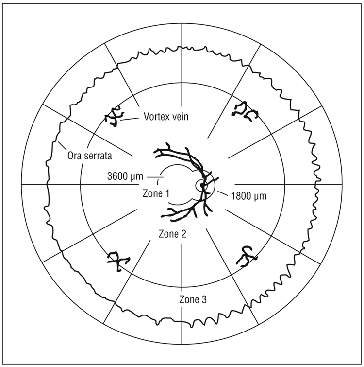
Retinal zonal topography, modified after Holland and colleagues. One disc diameter is approximately 1800 μm.

Proportionate zonal representation as estimated from both photographic and human anatomical studies.*Zones 1 and 2 from NTS accession No. PB97-192082. †Zone 3 from Curcio and Allen. ‡Rounded to the nearest fifth percentile.
Comment on
-
A controlled retrospective study of ganciclovir treatment for cytomegalovirus retinopathy. Use of a standardized system for the assessment of disease outcome. UCLA CMV Retinopathy. Study Group.Arch Ophthalmol. 1989 Dec;107(12):1759-66. doi: 10.1001/archopht.1989.01070020841024. Arch Ophthalmol. 1989. PMID: 2556989 Clinical Trial.
References
-
- Holland GN, Buhles WC Jr, Mastre B, Kaplan HJ; UCLA CMV Retinopathy Study Group. A controlled retrospective study of ganciclovir treatment for cytomegalovirus retinopathy: use of a standardized system for the assessment of disease outcome. Arch Ophthalmol. 1989;107(12):1759–1766. - PubMed
-
- Wei LL, Park SS, Skiest DJ. Prevalence of visual symptoms among patients with newly diagnosed cytomegalovirus retinitis. Retina. 2002;22(3):278–282. - PubMed
-
- Danis RP. The clinical site-reading center partnership in clinical trials. Am J Ophthalmol. 2009;148(6):815–817. - PubMed
-
- Studies of the Ocular Complications of AIDS Research Group. SOCA Cytomegalovirus Retinitis Grading Protocol. Springfield, VA: National Technical Information Service, US Department of Commerce; 1997. NTIS accession PB97-192082.
-
- Weinberg DV, Holbrook JT, Hubbard LD, Davis MD,Jabs DA, Holland GN; Studies of Ocular Complications of AIDS Research Group. Clinician versus reading center assessment of cytomegalovirus retinitis lesion size. Ophthalmology. 2005;112(4):559–566. - PubMed
