Review
. 2011 Nov 14;195(4):543-52.
doi: 10.1083/jcb.201103106.

The secret life of α-catenin: moonlighting in morphogenesis

Affiliations
Review

The secret life of α-catenin: moonlighting in morphogenesis

Stephanie L Maiden et al. J Cell Biol. .

Abstract

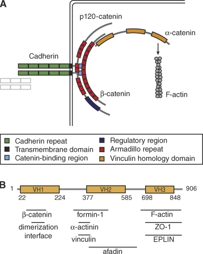
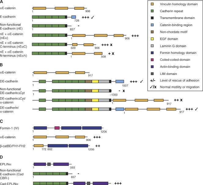
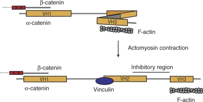
Cadherin-based intercellular adhesions are important determinants of proper tissue architecture. These adhesions must be both stable and dynamic to maintain tissue integrity as cells undergo morphogenetic movements during development. The role of α-catenin in this process has been vigorously debated due to conflicting in vitro and in vivo evidence regarding its molecular mechanism of action. Recent data supports the classical view that α-catenin facilitates actin attachments at adherens junctions, but also suggests that α-catenin may act as a force transducer, and may have additional roles in the cytoplasm. These multiple functions for α-catenin converge on the regulation of adhesion and may help to explain its stable yet dynamic nature.


Figures

Figure 1.
Figure 1.
The role of α-catenin in cell–cell adhesion. (A) The cadherin–catenin complex (CCC). The transmembrane cadherin mediates cell–cell adhesion through calcium-dependent homophilic binding of an adjacent cadherin. Intracellularly, cadherin interacts directly with p120-catenin and β-catenin. α-Catenin joins the complex by binding to β-catenin through its N terminus, while the C-terminal actin-binding domain recruits the actin cytoskeleton. (B) Key features of α-catenin, including the three vinculin homology (VH) domains and the binding sites of several proteins discussed in this review.
Figure 2.
Figure 2.
Structure and function of chimeric adhesion constructs. Fusions between the cytoplasmic tail of cadherins and α-catenin, or between cadherin and actin-binding proteins, have been used to probe the connection between the CCC and the actin cytoskeleton. Note: Not all of the chimeras used in each experiment are depicted in this schematic; only those discussed in this review are shown. (A) Chimeric fusion constructs of mammalian E-cadherin and αE-catenin (Nagafuchi et al., 1994; Imamura et al., 1999). (B) Chimeric fusions of Drosophila DE-cadherin and α-catenin (Pacquelet and Rørth, 2005). (C) Chimeric fusion of mammalian αE-catenin and formin-1(IV) (Kobielak et al., 2004). (D) Chimeric fusion of mammalian E-cadherin and EPLINα (Abe and Takeichi, 2008).
Figure 3.
Figure 3.
A model for tension-induced conformational changes in αE-catenin. αE-catenin may act as a mechanosensor to transduce changes in force to changes in the strength of cell–cell adhesions by recruiting additional adaptor proteins. See the text for further discussion. When αE-catenin is not under tension, its conformation may allow an inhibitory region to block its vinculin binding site. Upon the application of force, such as actomyosin-mediated contraction transduced through is C-terminal F-actin binding domain, αE-catenin may undergo a conformational change that displaces the inhibitory region from the vinculin binding site, allowing vinculin to bind. Vinculin, which has its own F-actin binding site, may in turn recruit additional F-actin to the CCC (Yonemura et al., 2010).
Figure 4.
Figure 4.
Non-junctional, homodimeric αE-catenin regulates membrane dynamics. In this model, there are two distinct populations of αE-catenin. At the AJ, αE-catenin associates with the CCC and mediates attachment to the actin cytoskeleton, either directly or indirectly. Near the membrane, a cytosolic pool of αE-catenin is created through dissociation from the CCC. This cytosolic pool has a high local concentration and can therefore form αE-catenin homodimers that are able to inhibit the Arp2/3 complex and thereby dampen membrane dynamics by preventing the formation of branched F-actin (Benjamin et al., 2010).

References

    1. Abe K., Takeichi M. 2008. EPLIN mediates linkage of the cadherin catenin complex to F-actin and stabilizes the circumferential actin belt. Proc. Natl. Acad. Sci. USA. 105:13–19 10.1073/pnas.0710504105 - DOI - PMC - PubMed
    1. Aberle H., Butz S., Stappert J., Weissig H., Kemler R., Hoschuetzky H. 1994. Assembly of the cadherin-catenin complex in vitro with recombinant proteins. J. Cell Sci. 107:3655–3663 - PubMed
    1. Anastasiadis P.Z. 2007. p120-ctn: A nexus for contextual signaling via Rho GTPases. Biochim. Biophys. Acta. 1773:34–46 10.1016/j.bbamcr.2006.08.040 - DOI - PubMed
    1. Barstead R.J., Waterston R.H. 1989. The basal component of the nematode dense-body is vinculin. J. Biol. Chem. 264:10177–10185 - PubMed
    1. Benjamin J.M., Nelson W.J. 2008. Bench to bedside and back again: molecular mechanisms of alpha-catenin function and roles in tumorigenesis. Semin. Cancer Biol. 18:53–64 10.1016/j.semcancer.2007.08.003 - DOI - PMC - PubMed

Publication types