Review
. 2011 Dec 27;366(1584):3605-24.
doi: 10.1098/rstb.2011.0069.

Structural insights into anaphase-promoting complex function and mechanism

Affiliations
Review

Structural insights into anaphase-promoting complex function and mechanism

David Barford. Philos Trans R Soc Lond B Biol Sci. .

Abstract

The anaphase-promoting complex or cyclosome (APC/C) controls sister chromatid segregation and the exit from mitosis by catalysing the ubiquitylation of cyclins and other cell cycle regulatory proteins. This unusually large E3 RING-cullin ubiquitin ligase is assembled from 13 different proteins. Selection of APC/C targets is controlled through recognition of short destruction motifs, predominantly the D box and KEN box. APC/C-mediated coordination of cell cycle progression is achieved through the temporal regulation of APC/C activity and substrate specificity, exerted through a combination of co-activator subunits, reversible phosphorylation and inhibitory proteins and complexes. Recent structural and biochemical studies of the APC/C are beginning to reveal an understanding of the roles of individual APC/C subunits and co-activators and how they mutually interact to mediate APC/C functions. This review focuses on the findings showing how information on the structural organization of the APC/C provides insights into the role of co-activators and core APC/C subunits in mediating substrate recognition. Mechanisms of regulating and modulating substrate recognition are discussed in the context of controlling the binding of the co-activator to the APC/C, and the accessibility and conformation of the co-activator when bound to the APC/C.


Figures

Figure 1.
Figure 1.
Primary structure of selective APC/C subunits. (a) Conserved TPR subunits of the APC/C: Cdc16, Cdc23, Cdc27 and Apc5. Schematic of Cdc27 and Cdc16 based on Zhang et al. [91,92], respectively. (b) Apc1 showing the PC (proteasome–cyclosome repeats). (c) Apc2 showing N-terminal cullin repeats and the C-terminal domain (CTD) responsible for binding Apc11. (d) Cdh1. ‘P’ indicates consensus Cdk-phosphorylation sites that are responsible for blocking APC/C interaction with Cdh1.
Figure 2.
Figure 2.
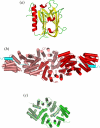
Crystal structures of selective APC/C subunits. (a) Apc10/Doc1 (S. cerevisiae) [102]. (b) Cdc16–Cdc26 heterodimer [91]. Cdc16 and Cdc27 share a related architecture and mode of homodimerization. (c) Cdc27 homodimer [91,92]. Both Cdc16 and Cdc27 homodimerize through an N-terminal domain composed of seven TPR units forming a TPR superhelix.
Figure 3.
Figure 3.
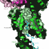
The N-terminal Met of Cdc26 is acetylated and enclosed within a chamber formed from the Cdc16 TPR superhelix and the homodimer interface. The N-terminal region of Cdc26 is shown in cyan with Cdc16 shown as a surface representation, with secondary structure indicated. Reproduced with permission from [91].
Figure 4.
Figure 4.
Comparison of the EM structure of the APC/C from S. cerevisiae, S. pombe and human APC/C shows similar overall structures. (a) Negative-stain EM map of S. cerevisiae APC/C [63] and (b) the cryo-EM map of S. cerevisiae APC/CCdh1–D box [63] are compared with (c) the cryo-EM map of S. pombe APC/C at 27 Å resolution [112] and (d) the cryo-negative-stain EM map of human APC/C at 25–19 Å resolution [111]. Stars denote positions of subunits identified by antibody labelling. Reproduced with permission from da Fonseca et al. [63].
Figure 5.
Figure 5.
Cryo-EM reconstruction of budding yeast APC/CCdh1–D box reveals the lattice-like architecture of the complex. Two views of the complex. Resolution is 10 Å.
Figure 6.
Figure 6.
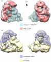
Three-dimensional electron microscopy structure comparisons of recombinant APC/C and APC/C subcomplexes. (a) Superimposition of TPR6 (red) onto endogenous APC/CCdh1 (blue mesh) and (b) SC8 (yellow) onto recombinant APC/C (purple mesh). Reproduced with permission from Schreiber et al. [81].
Figure 7.
Figure 7.
Three-dimensional localization of TPR subunits and atomic coordinate docking. (a) Three-dimensional localization of Cdc27–Apc9 by subtracting the APC/CΔCdc27ΔApc9 EM map from the recombinant APC/C map. The difference density is drawn as a grey mesh and used as restraints for Cdc27 docking. The two subunits within the homodimer are coloured in different shades of green. The symmetry axis of the Cdc27 homodimer is indicated in the right panel. (b) Three-dimensional localization of Cdc16–Cdc26–Apc13. The difference density (grey mesh) was calculated by subtracting the SC8 from the APC/CΔCdc27ΔApc9 EM map. The atomic coordinates of the S. pombe Cdc16–Cdc26 heterotetramer were used for rigid body docking. The two Cdc16 subunits within the heterotetramer are shown in red and light red and the Cdc26 N-terminus is shown in cyan. The molecular envelope corresponds to the APC/CΔCdc27ΔApc9 EM structure with density assigned to SC8 in yellow surface representation. The symmetry axis of the Cdc16–Cdc26 heterotetramer is indicated in the right panel. Reproduced with permission from Schreiber et al. [81].
Figure 8.
Figure 8.
Negative-stain EM reconstructions of S. cerevisiae APC/C show positions of Cdh1 and Apc10. Molecular envelopes of (a) APC/CCdh1, (b) apo APC/C, and (c) APC/CΔApc10–Cdh1. Density assigned to Cdh1 and Apc10 is shown in magenta and blue, respectively. The resolution of the APC/CCdh1 binary complex is 18–20 Å. Reproduced with permission from da Fonseca et al. [63].
Figure 9.
Figure 9.
Subunit organization and the pseudo-atomic model of APC/C. Atomic coordinates of Cdc16–Cdc26, Cdc23, Cdc27, Apc2, Apc10 and Cdh1 were docked in the 10 Å cryo-EM map of the APC/CCdh1–D box ternary complex represented in the grey mesh. The surface molecular boundaries of Apc1 (salmon) and Apc4–Apc5 (green) are indicated. Symmetry-related monomers of the Cdc16, Cdc23 and Cdc27 homodimers are represented in light and dark red, orange and green, respectively. Local twofold symmetry axes of Cdc27 and Cdc23 are indicated by diamonds. (a) View onto the central cavity orthogonal to the dyad axis of the Cdc27 homodimer. (b,c) Views related to (a) by rotations shown. (d) View approximately coincident with the Cdc16–Cdc26 dyad axis. Red spheres indicate the C-termini of Cdc16 and Cdc23, whereas red and blue spheres in Cdc27 denote the N- and C-termini of the inter-TPR insert. PC repeats of Apc1 are indicated. Reproduced with permission from Schreiber et al. [81].
Figure 10.
Figure 10.
Negative-stain EM reconstructions of the APC/C show that substrate binding to APC/CCdh1 involves Cdh1 and Apc10. Negative-stain EM reconstructions of (a) the S. cerevisiae APC/CCdh1–Hsl1 complex, (b) S. cerevisiae APC/CCdh1–D box, (c) S. cerevisiae APC/CCdh1–KEN box, (d) human APC/CCdh1 (Cdh1 in red), and (e) human APC/CCdh1–Hsl1 (Hsl1 in purple). Lower panels in (ac) show details of the structural changes associated with Cdh1 and Apc10 in the presence of substrate compared with the superimposed binary APC/CCdh1 map represented in the mesh. Panels (ac) reproduced with permission from da Fonseca et al. [63] and (d,e) reproduced with permission from Buschhorn et al. [113].
Figure 11.
Figure 11.
Cdh1, Apc10, Apc2 and Apc11 form a substrate-recognition catalytic module. (a) View of the cryo-EM APC/CCdh1–D box complex. Protein density is represented by a mesh with fitted atomic coordinates of the Cdh1 β-propeller (modelled), Apc10, Apc2–Apc11 and Cdc27. The two subunits of Cdc27 are shown in light and dark green. The view shows the twofold symmetry axis of Cdc27. Density connecting Cdh1 to a TPR superhelix of the Cdc27 dimer is indicated by an arrow. TPR motifs 8–10 of Cdc27, implicated in IR tail recognition [64], are shown in lighter colours. (b,c) Modelling of the D box peptide into density bridging Cdh1 and Apc10. (b) As an 8-residue α-helix. (c) As an extended chain. In both instances, the binding site for the D box is shared between Cdh1 and Apc10. (d) Schematic of the combined catalytic and substrate-recognition module responsible for D box binding and substrate ubiquitylation. D box is represented as binding to an interface between Cdh1 and Apc10. Reproduced with permission from da Fonseca et al. [63].
Figure 12.
Figure 12.
Docking of the pseudo-atomic model of S. cerevisiae APC/C into the electron microscopy-derived molecular envelope of the human APC/C–MCC complex reveals the position of the MCC and Apc7. Two views of the complex with the molecular envelope of human APC/C–MCC [111] are shown in mesh. Coordinates from Schreiber et al. [81]. Note the downward shift of the co-activator (here shown as Cdh1 in purple) when bound to the APC/C compared with the MCC.

References

    1. Irniger S., Piatti S., Michaelis C., Nasmyth K. 1995. Genes involved in sister chromatid separation are needed for B-type cyclin proteolysis in budding yeast. Cell 81, 269–27810.1016/0092-8674(95)90337-2 (doi:10.1016/0092-8674(95)90337-2) - DOI - DOI - PubMed
    1. King R. W., Peters J. M., Tugendreich S., Rolfe M., Hieter P., Kirschner M. W. 1995. A 20S complex containing CDC27 and CDC16 catalyzes the mitosis-specific conjugation of ubiquitin to cyclin B. Cell 81, 279–28810.1016/0092-8674(95)90338-0 (doi:10.1016/0092-8674(95)90338-0) - DOI - DOI - PubMed
    1. Sudakin V., Ganoth D., Dahan A., Heller H., Hershko J., Luca F. C., Ruderman J. V., Hershko A. 1995. The cyclosome, a large complex containing cyclin-selective ubiquitin ligase activity, targets cyclins for destruction at the end of mitosis. Mol. Biol. Cell 6, 185–197 - PMC - PubMed
    1. Tugendreich S., Tomkiel J., Earnshaw W., Hieter P. 1995. CDC27Hs colocalizes with CDC16Hs to the centrosome and mitotic spindle and is essential for the metaphase to anaphase transition. Cell. 81, 261–26810.1016/0092-8674(95)90336-4 (doi:10.1016/0092-8674(95)90336-4) - DOI - DOI - PubMed
    1. Peters J. M. 2006. The anaphase promoting complex/cyclosome: a machine designed to destroy. Nat. Rev. Mol. Cell Biol. 7, 644–65610.1038/nrm1988 (doi:10.1038/nrm1988) - DOI - DOI - PubMed

Publication types

Substances

LinkOut - more resources