. 2011 Dec 15;187(12):6171-5.
doi: 10.4049/jimmunol.1100835. Epub 2011 Nov 14.

Cutting edge: microRNA-181 promotes human NK cell development by regulating Notch signaling

Affiliations

Cutting edge: microRNA-181 promotes human NK cell development by regulating Notch signaling

Frank Cichocki et al. J Immunol. .

Abstract

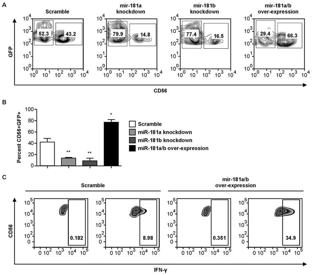
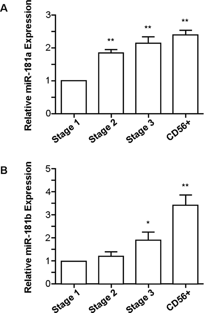
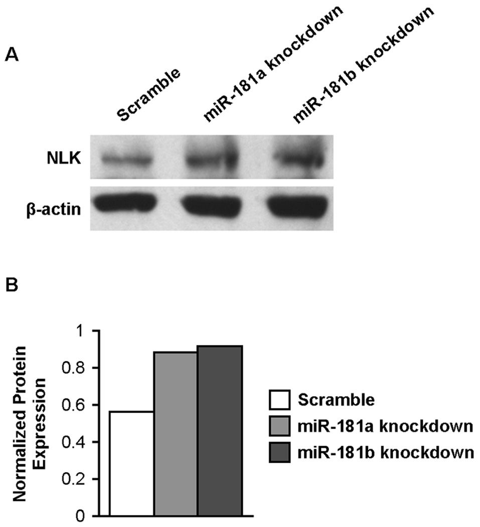
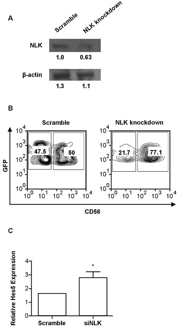
MicroRNAs (miRs) have recently been identified as important regulators of gene expression at the posttranscriptional level. Although it has clearly been established that miRs influence the ontogeny of several immune cell lineages, the role of individual miRs during NK cell development has not been described. In this study, we show that miR-181 expression levels have a profound impact on the development of human NK cells from CD34(+) hematopoietic progenitor cells and IFN-γ production in primary CD56(+) NK cells. We also demonstrate that nemo-like kinase (NLK), an inhibitor of Notch signaling, is a target of miR-181 in NK cells, and knockdown of NLK mirrors the developmental effect of miR-181 overexpression. We conclude that miR-181 promotes NK cell development, at least in part, through the suppression of NLK, providing an important link between miRs and Notch signaling.


Conflict of interest statement

The authors have no financial conflicts.

Figures

Figure 1
Figure 1. The expression levels of miR-181a and miR-181b determine the efficiency of human NK cell development from CD34+ progenitors and IFN-γ production by primary CD56+ peripheral blood NK cells
(A) Purified CD34+ cells were transduced with lentiviral vectors over-expressing scramble microRNA, miR-181a antisense knockdown RNA, miR-181b antisense knockdown RNA or full-length precursor miR-181a/b RNA and cultured for 28 days in NK cell differentiation conditions. Representative FACS plots of CD56 expression in GFP+ cells are shown. (B) Cumulative data showing CD56 expression in GFP+ cells at day 28 after each set of transductions. Results are shown as the mean CD56 expression of 4 donors in two separate experiments with 2 donors in each experiment with error bars representing SEM. Statistically significant differences from the scramble control were determined by one-way ANOVA (*p<0.05, **p<0.01). (C) GFP-gated FACS plots of intracellular IFN-γ production in CD56+ peripheral blood NK cells transduced with lentiviral vectors over-expressing scramble microRNA or full-length precursor miR-181a/b RNA and stimulated IL-12/18. Plots are representative of the analysis of three healthy donors.
Figure 2
Figure 2. The expression levels of miR-181a and miR-181b increase throughout NK cell development
qRT-PCR analysis of (A) miR-181a and (B) miR-181b levels throughout NK cell development (n=4). Results are shown as the mean expression values normalized against stage 1. Error bars represent SEM, and statistical significance was determined by one-way ANOVA (*p<0.05, **p<0.01).
Figure 3
Figure 3. Knockdown of miR-181a and miR-181b increase NLK protein levels in NK cells
(A) Western blots of NLK and β-actin protein in sort-purified NK92 cells transduced with lentiviral vectors over-expressing scramble microRNA, miR-181a antisense knockdown RNA or miR-181b antisense knockdown RNA. (B) Densitometric analysis of NLK protein expression normalized against β-actin protein levels for each lane. Results are representative of two independent experiments.
Figure 4
Figure 4. Knockdown of NLK promotes NK cell development and is associated with an increase in Hes5 expression
(A) Western blots of NLK and β-actin protein in sort-purified HEK293T cells transduced with scramble and siNLKlentiviral vectors. Values shown below each band represent denstometric analysis of protein expression. (B) Purified CD34+ cells were transduced with lentiviral vectors over-expressing scramble siRNA or NLK siRNA and cultured in NK cell differentiation conditions for 28 days. Representative FACS plots from GFP+ cells are shown. (C) qRT-PCR analysis of Hes5 expression in NK cells from scramble siRNA and NLK siRNA in GFP-isolated CD56+ cells from day 28 cultures (n=3). Error bars represent SEM, and statistical significance was determined by one-way ANOVA (*p<0.05).

References

    1. Bartel DP. MicroRNAs: genomics, biogenesis, mechanism, and function. Cell. 2004;116:281–297. - PubMed
    1. O’Connell RM, Rao DS, Chaudhuri AA, Baltimore D. Physiological and pathological roles for microRNAs in the immune system. Nat. Rev. Immunol. 2010;10:111–122. - PubMed
    1. O’Connell RM, Kahn D, Gibson WS, Round JL, Scholz RL, Chaudhuri AA, Kahn ME, Rao DS, Baltimore D. MicroRNA-155 promotes autoimmune inflammation by enhancing inflammatory T cell development. Immunity. 2010;33:607–619. - PMC - PubMed
    1. Xiao C, Calado DP, Galler G, Thai TH, Patterson HC, Wang J, Rajewsky N, Bender TP, Rajewsky K. MiR-150 controls B cell differentiation by targeting the transcription factor c-Myb. Cell. 2007;131:146–159. - PubMed
    1. Rao DS, O’Connell RM, Chaudhuri AA, Garcia-Flores Y, Geiger TL, Baltimore D. MicroRNA-34a perturbs B lymphocyte development by repressing the forkhead box transcription factor Foxp1. Immunity. 2010;33:48–59. - PMC - PubMed

Publication types

MeSH terms