. 2011 Nov 9:5:69.
doi: 10.3389/fnint.2011.00069. eCollection 2011.

Moving forward: advances in the treatment of movement disorders with deep brain stimulation

Affiliations

Moving forward: advances in the treatment of movement disorders with deep brain stimulation

Terry K Schiefer et al. Front Integr Neurosci. .

Abstract

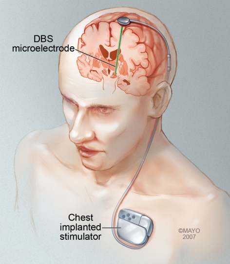
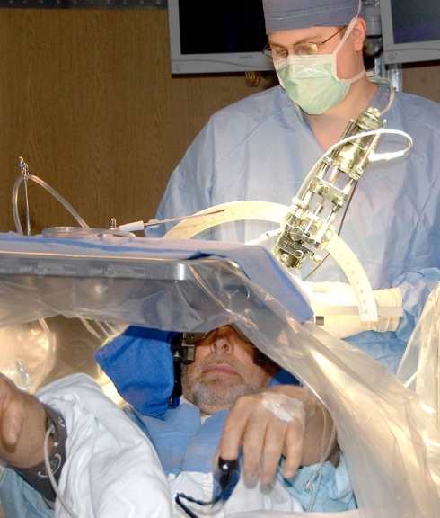
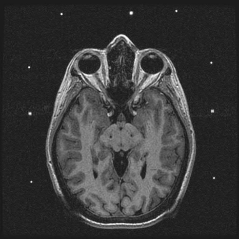
The modern era of stereotactic and functional neurosurgery has ushered in state of the art technologies for the treatment of movement disorders, particularly Parkinson's disease (PD), tremor, and dystonia. After years of experience with various surgical therapies, the eventual shortcomings of both medical and surgical treatments, and several serendipitous discoveries, deep brain stimulation (DBS) has risen to the forefront as a highly effective, safe, and reversible treatment for these conditions. Idiopathic advanced PD can be treated with thalamic, globus pallidus internus (GPi), or subthalamic nucleus (STN) DBS. Thalamic DBS primarily relieves tremor while GPi and STN DBS alleviate a wide range of Parkinsonian symptoms. Thalamic DBS is also used in the treatment of other types of tremor, particularly essential tremor, with excellent results. Both primary and various types of secondary dystonia can be treated very effectively with GPi DBS. The variety of anatomical targets for these movement disorders is indicative of the network-level dysfunction mediating these movement disturbances. Despite an increasing understanding of the clinical benefits of DBS, little is known about how DBS can create such wide sweeping neuromodulatory effects. The key to improving this therapeutic modality and discovering new ways to treat these and other neurologic conditions lies in better understanding the intricacies of DBS. Here we review the history and pertinent clinical data for DBS treatment of PD, tremor, and dystonia. While multiple regions of the brain have been targeted for DBS in the treatment of these movement disorders, this review article focuses on those that are most commonly used in current clinical practice. Our search criteria for PubMed included combinations of the following terms: DBS, neuromodulation, movement disorders, PD, tremor, dystonia, and history. Dates were not restricted.

Keywords: Parkinson’s disease; deep brain stimulation; dystonia; neuromodulation; tremor.


Figures

Figure 1
Figure 1
Illustration of a DBS system with a microelectrode implanted deep within the brain. This microelectrode is connected to a programmable stimulator/battery that is typically implanted in the chest in the subclavicular space. Permission to use this image granted by Mayo Clinic.
Figure 2
Figure 2
Intraoperative photo of a patient undergoing DBS surgery for Parkinson’s disease. As can be seen in the picture, patients are typically kept awake during the surgery to clinically assess the efficacy of electrode placement with real time stimulation, as well as any unwanted side effects. If the location is deemed suboptimal, it can easily be changed intraoperatively and reassessed.
Figure 3
Figure 3
Postoperative MRI demonstrating placement of electrodes in the STN bilaterally for treatment of idiopathic advanced PD.
Figure 4
Figure 4
Wireless instantaneous neurotransmitter concentration system (WINCS). (A) Photograph of the WINCS circuit board demonstrating the size, microprocessor, and Bluetooth components. (B) Photograph of WINCS sterilizable casing and the reference and working electrode leads. (C) Pseudocolor plot of stimulated dopamine in a pig with the Medtronic 3389 electrode in the STN and the WINCS carbon fiber in the caudate nucleus. The stimulation parameters are: 3 mA, 120 Hz, 2 s stim, 0.5 ms pulse width. The y-axis is the voltage cycled from −0.4 V at the top, to +1.5 V in the middle, and −0.4 V in the bottom; the x-axis is time. These scans occur 10 times per second and take 1/100 of a second. The scan parameters are cyclic, −0.4 to +1.5 to −0.4 V. The current is also recorded in nanoamps and shown by the color bar on the right. Inset at the upper right corner is the unfolded subtracted voltammogram. The oxidation peak is at +0.6 V which is typical for dopamine; the downward deflection is the reduction peak.

References

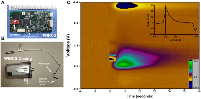
    1. Agnesi F., Tye S. J., Bledsoe J. M., Griessenauer C. J., Kimble C. J., Sieck G. C., Bennet K. E., Garris P. A., Blaha C. D., Lee K. H. (2009). Wireless instantaneous neurotransmitter concentration system-based amperometric detection of dopamine, adenosine, and glutamate for intraoperative neurochemical monitoring. J. Neurosurg. 111, 701–71110.3171/2009.3.JNS0990 - DOI - PMC - PubMed
    1. Alarcon F., Zijlmans J. C., Duenas G., Cevallos N. (2004). Post-stroke movement disorders: report of 56 patients. J. Neurol. Neurosurg. Psychiatr. 75, 1568–157410.1136/jnnp.2003.011874 - DOI - PMC - PubMed
    1. Alterman R. L., Miravite J., Weisz D., Shils J. L., Bressman S. B., Tagliati M. (2007). Sixty hertz pallidal deep brain stimulation for primary torsion dystonia. Neurology 69, 681–68810.1212/01.wnl.0000267430.95106.ff - DOI - PubMed
    1. Anderson V. C., Burchiel K. J., Hogarth P., Favre J., Hammerstad J. P. (2005). Pallidal vs subthalamic nucleus deep brain stimulation in Parkinson disease. Arch. Neurol. 62, 554–56010.1001/archneur.62.4.554 - DOI - PubMed
    1. Aybek S., Gronchi-Perrin A., Berney A., Chiuve S. C., Villemure J. G., Burkhard P. R., Vingerhoets F. J. (2007). Long-term cognitive profile and incidence of dementia after STN-DBS in Parkinson’s disease. Mov. Disord. 22, 974–98110.1002/mds.21478 - DOI - PubMed

LinkOut - more resources