

Splenic complications of sickle cell anemia and the role of splenectomy
- PMID: 22084706
- PMCID: PMC3200071
- DOI: 10.5402/2011/864257
Splenic complications of sickle cell anemia and the role of splenectomy
Abstract
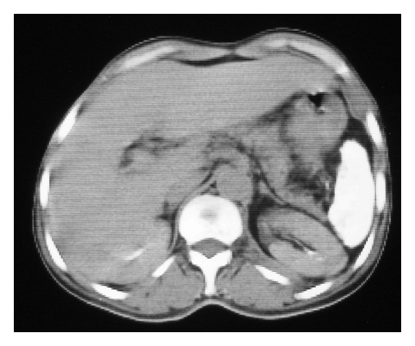
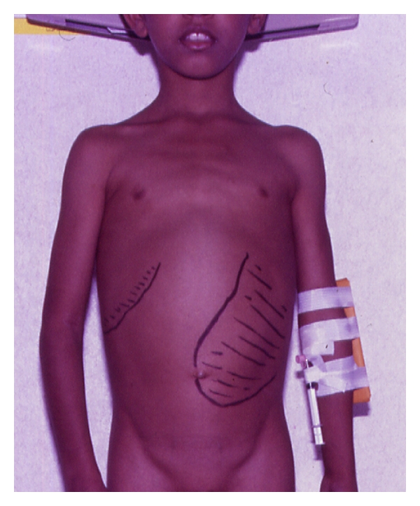
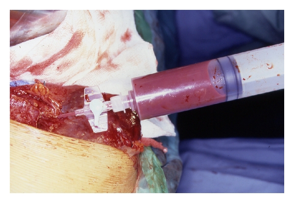
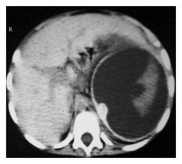
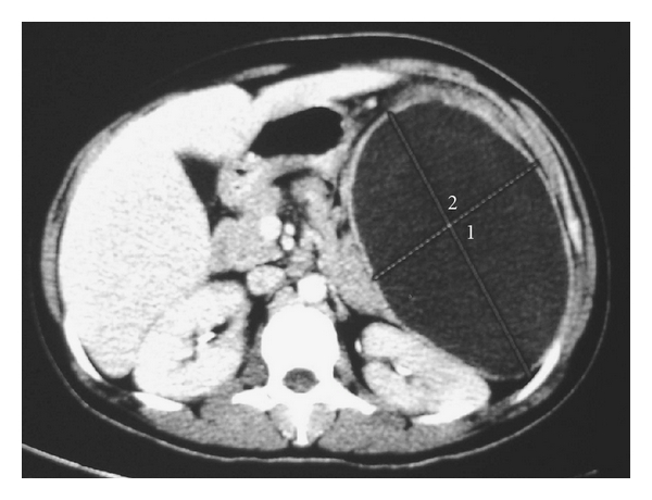
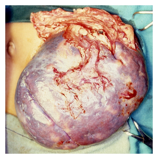
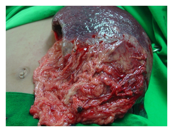
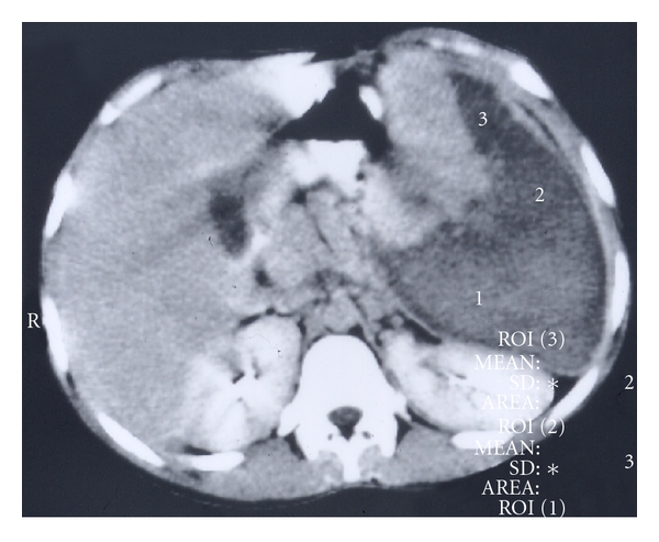
Sickle cell disease is one of the common hemoglobinopathies in the world. It can affect any part of the body and one of the most common and an early organ to be affected in SCA is the spleen. It is commonly enlarged during the first decade of life but then undergoes progressive atrophy leading to autosplenectomy. This however is not the case always and sometimes splenomegaly persist necessitating splenectomy for a variety of reasons including acute splenic sequestration crisis, hypersplenism, massive splenic infarction and splenic abscess. Splenic complications of SCA are known to be associated with an increased morbidity and in some it may lead to mortality. To obviate this, splenectomy becomes an essential part of their management. This review is based on our experience in the management of 173 children with various splenic complications of SCA necessitating splenectomy.
Figures
References
-
- Mason VR. Sickle cell anemia. Journal of the American Medical Association. 1922;79:1318–1320.
-
- Pauling L, Itano HA, Singer SJ, Wells IC. Sickle cell anemia, a molecular disease. Science. 1949;110(2865):543–548. - PubMed
-
- Ingram VM. A specific chemical difference between the globins of normal human and sickle-cell anæmia hæmoglobin. Nature. 1956;178(4537):792–794. - PubMed
-
- Ingram VM. Gene mutations in human hæmoglobin: the chemical difference between normal and sickle cell hæmoglobin. Nature. 1957;180(4581):326–328. - PubMed
LinkOut - more resources
Full Text Sources
