

Dysphagia lusoria: a case of an aberrant right subclavian artery and a bicarotid trunk
- PMID: 22084776
- PMCID: PMC3195806
- DOI: 10.5402/2011/819295
Dysphagia lusoria: a case of an aberrant right subclavian artery and a bicarotid trunk
Abstract
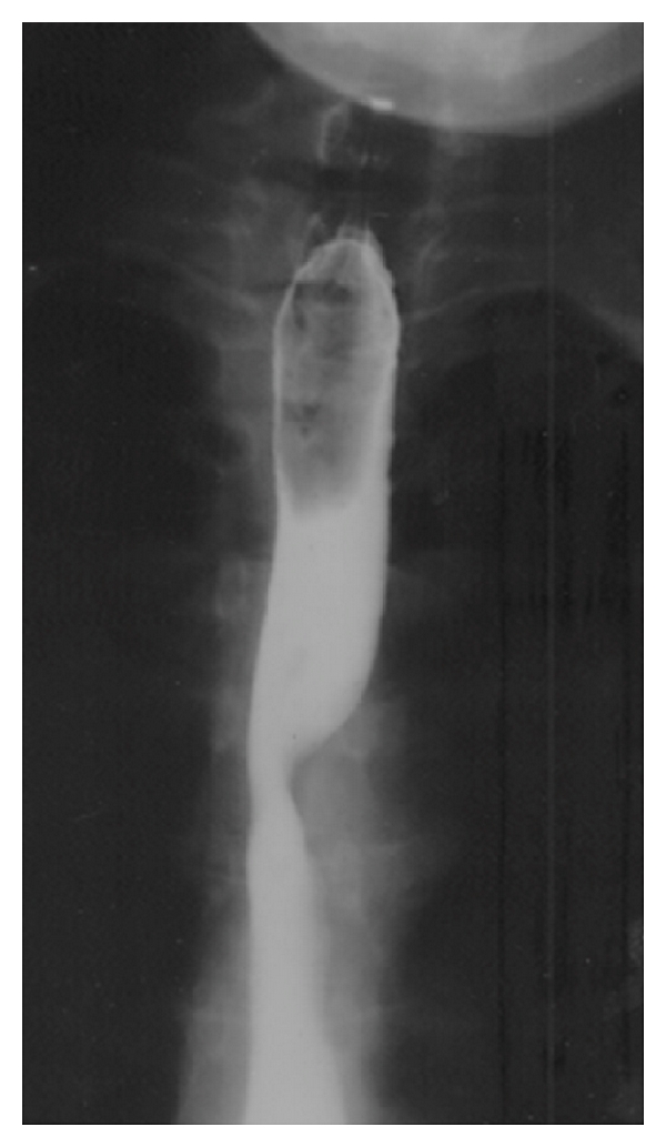
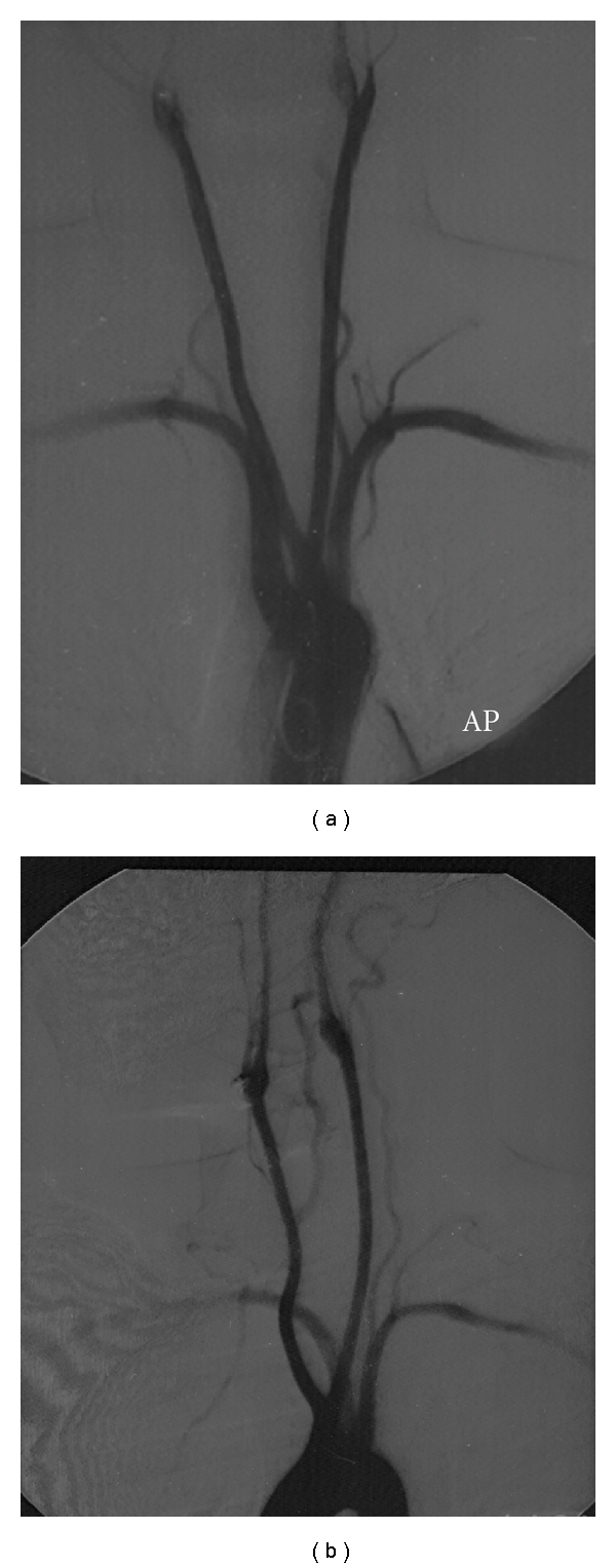
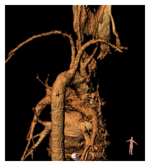
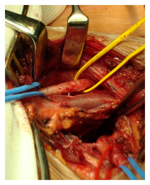
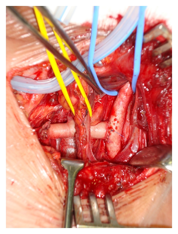
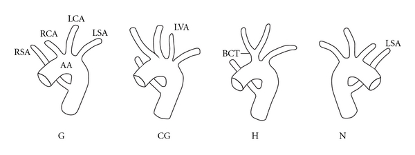
Dysphagia Lusoria is dysphagia secondary to an aberrant right subclavian artery that has a retroesophageal course. Adachi and Williams categorized aortic arch anomalies, showing that the right subclavian artery arising in this fashion (as the final branch of the descending aortic arch) is one of the more common. However, this very rarely coexists with a bicarotid trunk. We present such a case as it is manifested in a 36-year-old lady complaining of marked weight loss and dysphagia. The diagnosis remained elusive until a CT scan of the chest was performed; angiography further delineated the pathology. It is believed that the combination of the common carotid origins with the retroesophageal course of the aberrant vessel more frequently accounts for symptoms in the absence of an aneurysm of the origin of the aberrant vessel. Several techniques to manage the aberrant vessel have been described in the literature, but we favoured open ligation and transposition to the right carotid artery.
Figures
References
-
- Williams GD, Aff HM, Schmeckebier M, Edmonds HW, Grand EG. Variations in the arrangement of the branches arising from the aortic arch in the American whites and negroes. The Anatomical Record. 1932;54:247–251.
-
- Bayford D. An account of a singular case of obstructed degluitition. Memoirs of the Medical Society of London. 1794;2:275–286.
-
- Kommerell B. Verlagerung des Osophagus durch eine abnorm verlaufende Arteria subclavia dextra (Arteria lusoria) Fortschr Geb Roentgenstrahlen. 1936;54:590–595.
-
- Rahman HA, Sakurai A, Dong K, Setsu T, Umetani T, Yamadori T. The retroesophageal subclavian artery–a case report and review. Kaibogaku zasshi. Journal of anatomy. 1993;68(3):281–287. - PubMed
-
- Adachi B. Das Arteriensystem der Japener. Tokyo, Japan: Kenkyusha Press; 1928.
Publication types
LinkOut - more resources
Full Text Sources