

Review
doi: 10.5732/cjc.011.10213.
Epub 2011 Nov 15.
Schwannoma of the conus medullaris: a rare case
Affiliations
- PMID: 22085527
- PMCID: PMC4013335
- DOI: 10.5732/cjc.011.10213
Item in Clipboard
Review
Schwannoma of the conus medullaris: a rare case
Chin J Cancer.
2011 Dec.
Abstract
Intradural schwannoma of the conus medullaris is a rare form of spinal neoplasm, which commonly occurs in the lumbar region. Conus medullaris level is unusual for schwannomas. A 49-year-old woman presented with chronic sciatica, mild bladder dysfunction, and paresthesia in the buttocks. Magnetic resonance imaging of the spine showed a mass lesion in the conus medullaris region with nerve compression. The tumor was completely resected and diagnosed histologically as schwannoma. The patient recovered after surgery. Clinical and radiologic features of this rare tumor are reviewed and are accompanied by literature findings.
Figures
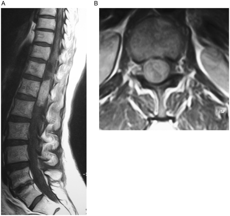
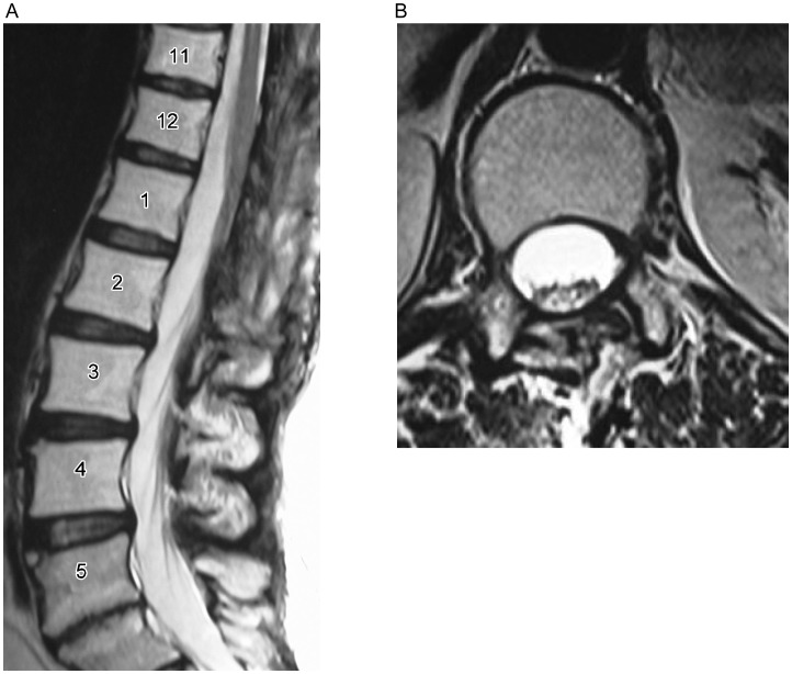
A, the sagittal T1-weighted MR image shows an intradural, isointense lesion of the conus medullaris. B, on the axial T1-weighted MR images, this lesion was heterogeneously enhanced with contrast. The spinal nerves are displaced laterally.
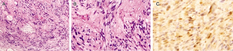
A, observing under a microscope, the tumor shows cellular and hipocellular areas (HE ×100). B, plumped spindle-shaped cells with palisading nuclei can be seen (HE ×100). C, S-100 immunoreactivity is observed in tumor cells (S-100 ×200).
References
-
- Asahara H, Kawai A, Harada Y, et al. Spinal schwannomas: a review of 42 cases [J] Acta Med Okayama. 1996;50(1):25–28. - PubMed
-
- Conti P, Pansini G, Mouchaty H, et al. Spinal neurinomas: retrospective analysis and long-term outcome of 179 consecutively operated cases and review of the literature [J] Surg Neurol. 2004;61(1):34–43. - PubMed
-
- Hasegawa M, Fujisawa H, Hayashi Y, et al. Surgical pathology of spinal schwannomas: a light and electron microscopic analysis of tumor capsules [J] Neurosurgery. 2001;49(6):1388–1393. - PubMed
-
- Jinnai T, Koyama T. Clinical characteristics of spinal nerve sheath tumors: analysis of 149 cases [J] Neurosurgery. 2005;56(3):510–515. - PubMed
-
- Celli P. Treatment of relevant nerve roots involved in nerve sheath tumors: removal or preservation? [J] Neurosurgery. 2002;51(3):684–692. - PubMed
Publication types
MeSH terms
Substances
LinkOut - more resources
Full Text Sources