. 2012 Feb;26(2):832-41.
doi: 10.1096/fj.11-189415. Epub 2011 Nov 15.

Oxidation of HRas cysteine thiols by metabolic stress prevents palmitoylation in vivo and contributes to endothelial cell apoptosis

Affiliations

Oxidation of HRas cysteine thiols by metabolic stress prevents palmitoylation in vivo and contributes to endothelial cell apoptosis

Joseph R Burgoyne et al. FASEB J. 2012 Feb.

Abstract

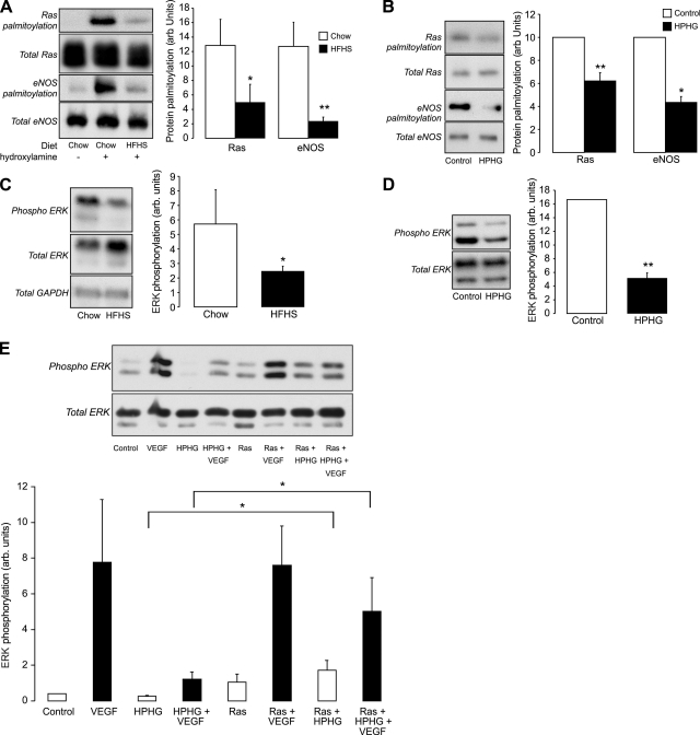
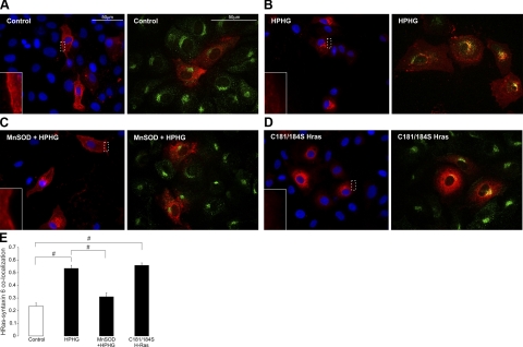
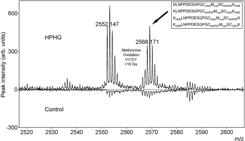
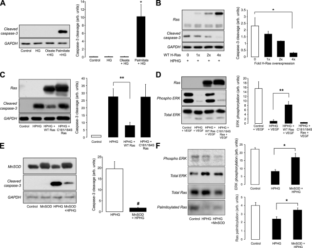
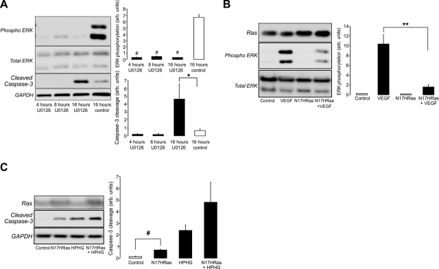
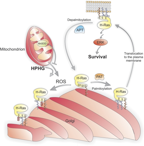
Here we demonstrate a new paradigm in redox signaling, whereby oxidants resulting from metabolic stress directly alter protein palmitoylation by oxidizing reactive cysteine thiolates. In mice fed a high-fat, high-sucrose diet and in cultured endothelial cells (ECs) treated with high palmitate and high glucose (HPHG), there was decreased HRas palmitoylation on Cys181/184 (61±24% decrease for cardiac tissue and 38±7.0% in ECs). This was due to oxidation of Cys181/184, detected using matrix-assisted laser desorption/ionization time of flight (MALDI TOF)-TOF. Decrease in HRas palmitoylation affected its compartmentalization and Ras binding domain binding activity, with a shift from plasma membrane tethering to Golgi localization. Loss of plasma membrane-bound HRas decreased growth factor-stimulated ERK phosphorylation (84±8.6% decrease) and increased apoptotic signaling (24±6.5-fold increase) after HPHG treatment that was prevented by overexpressing wild-type but not C181/184S HRas. The essential role of HRas in metabolic stress was made evident by the similar effects of expressing an inactive dominant negative N17-HRas or a MEK inhibitor. Furthermore, the relevance of thiol oxidation was demonstrated by overexpressing manganese superoxide dismutase, which improved HRas palmitoylation and ERK phosphorylation, while lessening apoptosis in HPHG treated ECs.


Figures

Figure 1.
Figure 1.
Effect on protein palmitoylation and growth factor signaling in BAECs treated with HPHG and hearts of mice fed HFHS diet. A) Ras (total Ras antibody) and eNOS palmitoylation is lower in cardiac tissue from mice fed a HFHS diet for 4 mo compared with a chow diet, as detected using the hydroxylamine dependent biotin-switch assay (n=4). B) Ras (total Ras antibody) and eNOS palmitoylation is also decreased in BAECs treated with medium containing HPHG for 16 h compared with control medium (n=6). C) In animals fed an HFHS diet, there is a substantial loss in basal ERK phosphorylation compared with mice fed chow (n=4). D) In BAECs treated with HPHG, there is also decreased basal ERK phosphorylation compared with control medium-treated cells (n=6). E) Basal and VEGF-induced ERK phosphorylation that was attenuated by preincubation of BAECs with HPHG was prevented in cells that overexpressed wild-type HRas (n=6). All graphs have been normalized for protein loading. Error bars = se. *P < 0.05, **P < 0.01; Student's t test.
Figure 2.
Figure 2.
Localization of HRas (Xpress-tag antibody, shown in red) in control medium or HPHG-treated BAECs. Left panels: fluorescence microscopy images of overexpressed HRas localization with DAPI nuclear staining in BAECs, including a magnified region of HRas plasma membrane staining. Right panels: confocal images showing colocalization of overexpressed HRas (Xpress-tag antibody, red) and the Golgi marker syntaxin-6 (green) in BAECs. A) In control medium, endothelial cell HRas is homogenously distributed throughout much of the cell and is also found at the plasma membrane. B) In BAECs treated with HPHG, HRas displays very little localization at the plasma membrane and instead is mostly found at the Golgi, which is evident from a high level of colocalization with syntaxin-6. C) Overexpression of MnSOD in BAECs before HPHG treatment prevents, to some extent, the redistribution of HRas by improving plasma membrane and cytosolic localization while decreasing that which is located at the Golgi. D) Overexpressed C181/184S HRas displays very little plasma membrane staining and instead colocalizes to a high extent with syntaxin-6 and also localizes around the outside of the nucleus. E) Quantification of the colocalization of HRas with the Golgi marker syntaxin-6 shows increased colocalization in BAECs treated with HPHG that is reversed by overexpression of MnSOD. Colocalization ranges between −1 for perfect exclusion and 1 for maximum correlation (n=20–25). Error bars = se. #P < 0.005; Student's t test.
Figure 3.
Figure 3.
Intracellular activity of HRas in treated BAECs. This was determined by analyzing the colocalization of RBD with HRas overexpressed (Xpress-tag antibody) in BAECs that had been stimulated for 5 min with VEGF before being fixed. A) In control medium-treated cells, there is some colocalization of HRas with RBD at the plasma membrane (indicated by the white arrows), which is also apparent in the fluorescence intensity scans generated along the white lines indicated for each analyzed cell. B) In HPHG-treated BAECs, there is less membrane colocalization and also no increase in RBD staining with the accumulated HRas at the Golgi as indicated in the intensity scan for each analyzed cell.
Figure 4.
Figure 4.
MS analysis of HRas from BAECs treated with control or HPHG medium. Comparison between the MALDI TOF spectra for overexpressed HRas purified from the cell lysate of BAECs treated with either control or HPHG medium after having been processed by the hydroxylamine dependent biotin-switch assay. In the spectrum for HRas from HPHG-treated cells, there were 2 peaks of interest that were more abundant than in the control group that had masses which equated to HNE, methionine oxidized and S-glutathiolated C-terminal fragments of HRas. MALDI TOF/TOF spectra of these peaks (Supplemental Fig. S2) revealed a complex mixture of oxidized HRas where either Cys181 or 184 was S-glutathiolated, with the other cysteine being palmitoylated (biotin-HPDP). In addition, Lys168 or 185 was modified by HNE, and in the right hand peaks, Met182 was also oxidized. Inset: various modifications of the C-terminal peptide identified using MALDI TOF/TOF in the highlighted peak (solid black arrow) of HRas from BAECs treated with HPHG.
Figure 5.
Figure 5.
Effect of HPHG on caspase-3 cleavage in treated BAECs. A) Treatment of BAECs with HG or HG with 0.4 mM of the unsaturated fatty acid oleate did not induce caspase-3 cleavage. However, treatment of BAECs with HF and 0.4 mM palmitate increased accumulation of this apoptotic signaling marker (n=3). B) Wild-type HRas overexpression (shown using total Ras antibody) prevented caspase-3 cleavage in BAECS treated with HPHG in a titer-dependent manner (n=3). C) Although the wild-type HRas prevented caspase-3 cleavage, the C181/184S was unable to protect cells from the apoptotic effect of HPHG (n=6). D) In BAECs, VEGF-dependent ERK phosphorylation was inhibited by HPHG, an effect that was prevented by overexpressing wild-type HRas but not the C181/184S mutant (n=3). E) Overexpression of the mitochondrial form of SOD, MnSOD, prevented caspase-3 cleavage in BAECs treated with HPHG (n=3). F) In BAECs overexpressing MnSOD and treated with HPHG, there is increased basal ERK phosphorylation and HRas palmitoylation compared with HPHG treatment alone (variations in amount of input protein is due to protein loss during multiple sample desalting steps during the biotin switch assay, n=3–4). All graphs have been normalized for protein loading. Error bars = se. *P < 0.05, **P < 0.01, #P < 0.005; Student's t test.
Figure 6.
Figure 6.
Effect of ERK inhibition and blocking HRas plasma membrane signaling in BAECs on growth factor and apoptotic signaling. A) Inhibition of MEK by U0126 attenuated basal ERK phosphorylation and increased caspase-3 cleavage 16 h after treatment. B) Overexpression of inactive dominant negative N17-HRas (total Ras antibody) in BAECs attenuated VEGF-dependent ERK phosphorylation (n=3). C) Overexpression of N17-HRas also increased cell apoptotic signaling in control medium-treated BAECs and did not protect cells from the apoptotic effect of HPHG (n=3). All graphs have been normalized for protein loading. Error bars = se. *P < 0.05, **P < 0.01, #P < 0.005; Student's t test.
Figure 7.
Figure 7.
Proposed model for the regulation of HRas palmitoylation under physiological conditions and in the presence of HPHG. HRas that has been processed by being sequentially farnesylated, proteolysed, and carboxyl methylated is palmitoylated at Cys181 and Cys184 at the Golgi membrane by the palmitoyl acyltransferase (PAT) under physiological conditions. Palmitoylation of HRas at both sites induces exocytic translocation of this GTPase from the Golgi to the plasma membrane, where it mediates growth factor signaling, including regulating the ERK pathway that is involved in endothelial cell survival. At the plasma membrane, HRas is depalmitoylated by the acyl protein thioesterase (APT), which causes the GTPase to return to the Golgi, where it can then be repalmitoylated and begin the transport cycle again. This cycle is disrupted by reactive oxygen species (ROS) generated by mitochondria, which are elevated by HPHG and can be attenuated by increased MnSOD activity. The increased ROS from the mitochondria in endothelial cells treated with HPHG oxidize either Cys181 or Cys184 on HRas, leading to S-glutathiolation on either cysteine, preventing palmitoylation at these sites. This results in the formation of monopalmitoylated HRas, which becomes trapped at the Golgi, as single-site palmitoylation is insufficient to mediate efficient exocytic transport to the plasma membrane.

References

    1. Wright L. P., Philips M. R. (2006) Thematic review series: lipid posttranslational modifications. CAAX modification and membrane targeting of Ras. J. Lipid Res. 47, 883–891 - PubMed
    1. Rocks O., Peyker A., Kahms M., Verveer P. J., Koerner C., Lumbierres M., Kuhlmann J., Waldmann H., Wittinghofer A., Bastiaens P. I. (2005) An acylation cycle regulates localization and activity of palmitoylated Ras isoforms. Science 307, 1746–1752 - PubMed
    1. Hancock J. F., Paterson H., Marshall C. J. (1990) A polybasic domain or palmitoylation is required in addition to the CAAX motif to localize p21ras to the plasma membrane. Cell 63, 133–139 - PubMed
    1. Swarthout J. T., Lobo S., Farh L., Croke M. R., Greentree W. K., Deschenes R. J., Linder M. E. (2005) DHHC9 and GCP16 constitute a human protein fatty acyltransferase with specificity for H- and N-Ras. J. Biol. Chem. 280, 31141–31148 - PubMed
    1. Duncan J. A., Gilman A. G. (1998) A cytoplasmic acyl-protein thioesterase that removes palmitate from G protein alpha subunits and p21(RAS). J. Biol. Chem. 273, 15830–15837 - PubMed

Publication types

MeSH terms