. 2011 Nov 15:11:332.
doi: 10.1186/1471-2148-11-332.

Emergence and evolution of the glycoprotein hormone and neurotrophin gene families in vertebrates

Affiliations

Emergence and evolution of the glycoprotein hormone and neurotrophin gene families in vertebrates

Sandra Dos Santos et al. BMC Evol Biol. .

Abstract

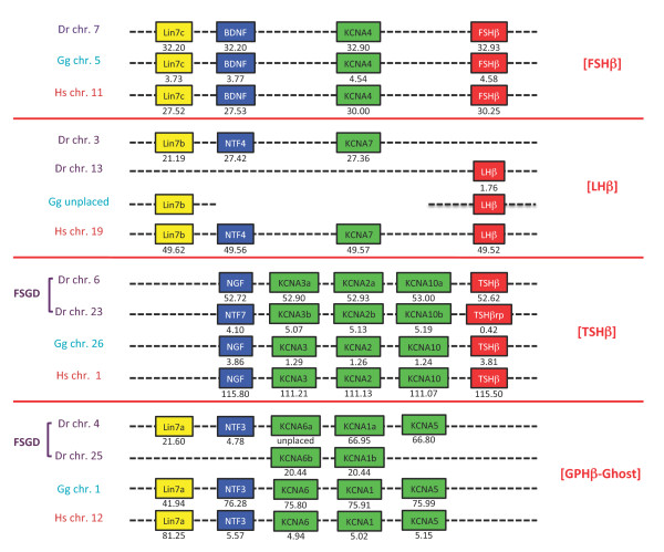
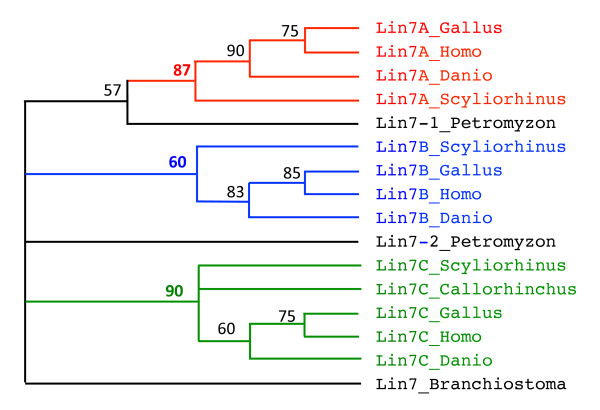
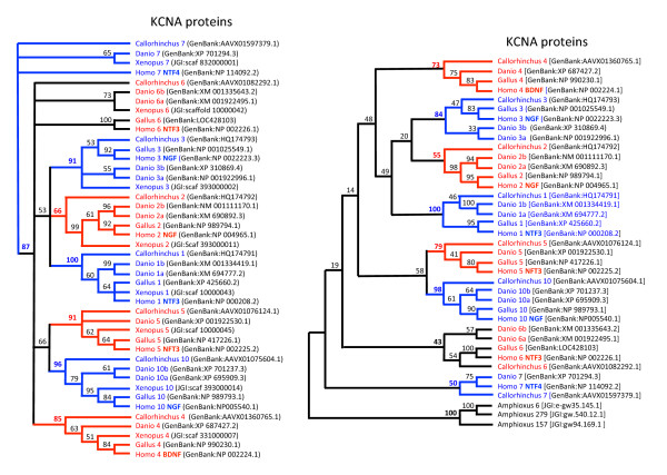
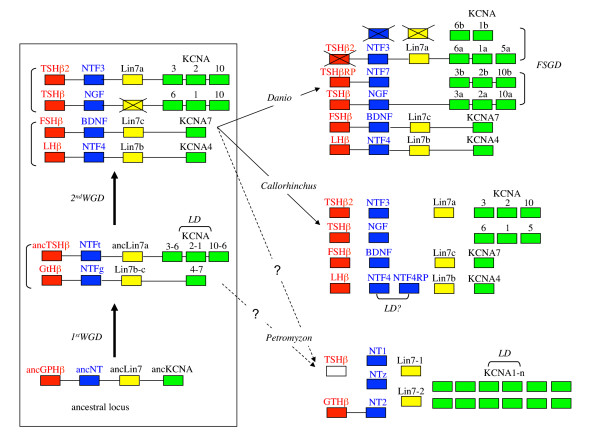
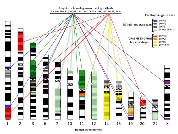
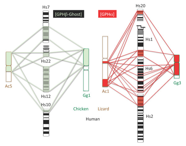
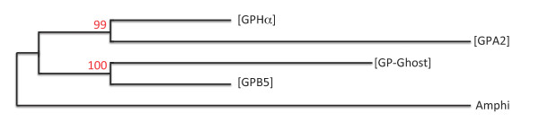
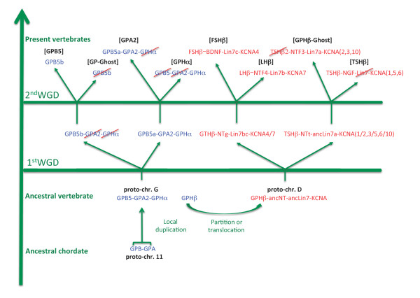
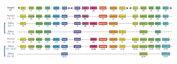
Background: The three vertebrate pituitary glycoprotein hormones (GPH) are heterodimers of a common α and a specific β subunit. In human, they are located on different chromosomes but in a similar genomic environment. We took advantage of the availability of genomic and EST data from two cartilaginous fish species as well as from two lamprey species to identify their repertoire of neurotrophin, lin7 and KCNA gene family members which are in the close environment of gphβ. Gphα and gphβ are absent outside vertebrates but are related to two genes present in both protostomes and deuterostomes that were named gpa2 and gpb5. Genomic organization and functional characteristics of their protein products suggested that gphα and gphβ might have been generated concomitantly by a duplication of gpa2 and gpb5 just prior to the radiation of vertebrates. To have a better insight into this process we used new genomic resources and tools to characterize the ancestral environment before the duplication occurred.

Results: An almost similar repertoire of genes was characterized in cartilaginous fishes as in tetrapods. Data in lampreys are either incomplete or the result of specific duplications and/or deletions but a scenario for the evolution of this genomic environment in vertebrates could be proposed. A number of genes were identified in the amphioxus genome that helped in reconstructing the ancestral environment of gpa2 and gpb5 and in describing the evolution of this environment in vertebrates.

Conclusion: Our model suggests that vertebrate gphα and gphβ were generated by a specific local duplication of the ancestral forms of gpa2 and gpb5, followed by a translocation of gphβ to a new environment whereas gphα was retained in the gpa2-gpb5 locus. The two rounds of whole genome duplication that occurred early in the evolution of vertebrates generated four paralogues of each gene but secondary gene losses or lineage specific duplications together with genomic rearrangements have resulted in the present organization of these genes, which differs between vertebrate lineages.


Figures

Figure 1
Figure 1
Comparative genomic environment of gphβ subunit genes in representative vertebrates. Human (Hs), chicken (Gg) and zebrafish (Dr) gphβ (lhβ, fshβ and tshβ) environment ([GPHβ]) are shown with their coordinates in megabase pairs from the p end of the relevant chromosome (chr.). [LHβ] is not found in chicken genomic databases but the existence of at least lhβ and lin7b is known from their cDNA product. A fish-specific genomic duplication (FSGD) generated specific duplicates in zebrafish. A paralogous environment devoid of any gphβ subunit gene is named [GPHβ-Ghost].
Figure 2
Figure 2
Phylogeny of Lin7b sequences. Phylogenetic reconstruction of chicken (Gallus), human (Homo), zebrafish (Danio) dogfish (Scyliorhinus), lamprey (Petromyzon) and amphioxus (Branchiostoma) lin7-related sequences was inferred using the maximum parsimony method (heuristic search). Bootstrapping (values at the nodes) was used over 1000 replicates. Lin7-A, -B and -C are clustered into monophyletic groups (bootstrap value in bold) that are highlighted in different colors.
Figure 3
Figure 3
Phylogeny of KCNA sequences. Left panel: Xenopus, chicken (Gallus), zebrafish (Danio), elephant shark (Callorhinchus) and human (Homo) KCNA sequences. Phylogenetic reconstructions were inferred using the maximum parsimony method (heuristic search). Bootstrapping (values at the nodes) was used over 1000 replicates. The KCNA type is indicated by the number following the species name. The neurotrophin-type to which they are neighbor in human genomes is indicated in bold with the human sequence reference. All but KCNA6 were included into monophyletic groups (highlighted by alternate colors) that were supported by bootstrap values of 50% and over (values in bold). Right panel: amphioxus sequences are substituted to Xenopus sequences. None of the amphioxus sequence is closer to either vertebrate KCNA type. Lower bootstrap values were obtained when amphioxus sequences were included because only truncated sequences could reliably be aligned.
Figure 4
Figure 4
Model for the evolution of gphβ/nt locus in vertebrates. Left panel: evolution of the ancestral locus with the first and second whole genome duplication (WGD). Right panel: resulting known genomic loci in sea lamprey Petromyzon marinus, elephant shark Callorhinchus milii and zebrafish Danio rerio. It is still to be established whether cyclostomes have been submitted to the two rounds of genome duplication or not. Duplicate loci resulting from WGD or the Fish Specific Genome Duplication (FSGD) are indicated by vertical brackets whereas horizontal brackets represent local gene duplications (LD). Crossed boxes represent genes that have been lost after duplication. Genes demonstrated to be gathered in a definite locus are linked by a horizontal bar. The origin of tshβ2 in Callorhinchus is uncertain, either resulting from the second round of WGD or from a local duplication of tshβ. A tshβ-type subunit gene is expected in lamprey but has not been characterized yet and is represented as an empty box. NTFt and NTFg are given symbols for ancestral NTs associated with TSH and GTH, respectively.
Figure 5
Figure 5
Gph-related gene-containing paralogous gene sets in human and amphioxus genomes. This figure is a schematic representation of the data presented in Additional files 7 and 9 that shows the genomic distribution of the paralogous gene sets (tetra-paralogons) containing the gph-related genes in the human genome and lists the most important scaffolds (genome version 2) where the amphioxus homologues are located. The scaffold V2_158 contains genes that are homologous to genes belonging to one or the other tetra-paralogon.
Figure 6
Figure 6
[GPHα] and [GPHβ -ghost]-containing paralogous gene sets in human, chicken and lizard genomes. Discrete gene locations are linked by dotted lines whereas more important loci are bordered by continuous lines (see Additional files 7 and 9 for details). The human chr. 1 was truncated for convenience. Hs: human; Gg: chicken; Ac: lizard.
Figure 7
Figure 7
Phylogenetic relationships between paralogous gene sets. Maximum Likelihood inferred relationships between concatenated protein sequences (1959 informative positions retained) of human RTN, MAP3K, MAP4K, ACTN, MARK, SIPA, KLC and FOS paralogues from the four ([GPHα], [GPB5], [GPA2] and [GP_Ghost]) paralogous gene sets with their homologue in amphioxus (Amphi) as the outgroup. Bootstrap values (100 replicates) are indicated in red.
Figure 8
Figure 8
Scenario for the evolution of the gph-related gene environments. The chordate gpa-gpb locus was duplicated prior to the first round of genomic duplication and the newly created gphβ was transferred to vertebrate proto-chr D whereas gpa2, gpb5 and gphα localized on vertebrate proto-chr. G. Paralogues of gpa2-gpb5-gphα and gphβ environments were then created through two whole genome duplications (WGD). Genes that have been lost are crossed out in red. Gpb5a is still present in teleosts in [GPA2] environment but was lost in tetrapods (see Figure 9).
Figure 9
Figure 9
Comparative genomic environment of gpa2 and gpb5. Genes are given with, underneath, their position in megabase pairs from the p end of each chromosome (chr.) in human and zebrafish (zebra) (see Additional file 10 for details and links to Ensembl website). Amphioxus (Amphi) homologues are given with the scaffold number (version 2) on which they are located. Names are after Ensembl in the human genome except for amphioxus homologues, which were reduced. Duplicated chromosome fragments in zebrafish are indicated with brackets. Due to rearrangements, the zebrafish gpb5b environment is scattered among chr. 13 and 17 whereas its FSGD driven duplicated environment is on chr. 20. Human GPB5 is orthologous to zebrafish GPB5b, whereas zebrafish GPB5a environment is orthologous to that of human GPA2.

References

    1. Quérat B, Sellouk A, Salmon C. Phylogenetic analysis of the vertebrate glycoprotein hormone family including new sequences of sturgeon Acipenser baeri, β-subunits of the two gonadotropins and the thyroid-stimulating hormone. Biol Reprod. 2000;63:222–228. doi: 10.1095/biolreprod63.1.222. - DOI - PubMed
    1. Quérat B, Tonnerre-Doncarli C, Géniès F, Salmon C. Duality of gonadotropins in gnathostomes. Gen Comp Endocrinol. 2001;124:308–314. doi: 10.1006/gcen.2001.7715. - DOI - PubMed
    1. Sower SA, Moriyama S, Kasahara M, Takahashi A, Nozaki M, Uchida K, Dahlstrom JM, Kawauchi K. Identification of sea lamprey GtHβ-like cDNA and its evolutionary implications. Gen Comp Endocrinol. 2006;148:22–32. doi: 10.1016/j.ygcen.2005.11.009. - DOI - PubMed
    1. Uchida K, Moriyama S, Chiba H, Shimotani T, Honda K, Miki M, Takahashi A, Sower SA, Nozaki M. Evolutionary origin of a functional gonadotropin in the pituitary of the most primitive vertebrate, hagfish. Proc Natl Acad Sci USA. 2010;107:15832–15837. doi: 10.1073/pnas.1002208107. - DOI - PMC - PubMed
    1. Huang EJ, Reichardt LF. Neurotrophins: roles in neuronal development and function. Annu Rev Neurosci. 2001;24:677–736. doi: 10.1146/annurev.neuro.24.1.677. - DOI - PMC - PubMed

Publication types