

Mammary gland morphological and gene expression changes underlying pregnancy protection of breast cancer tumorigenesis
- PMID: 22085904
- PMCID: PMC4116391
- DOI: 10.1152/physiolgenomics.00056.2011
Mammary gland morphological and gene expression changes underlying pregnancy protection of breast cancer tumorigenesis
Abstract
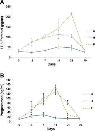
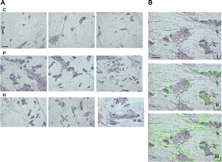
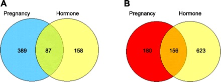
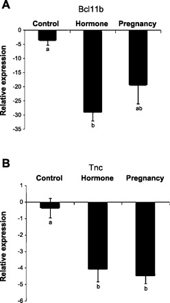
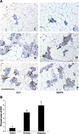
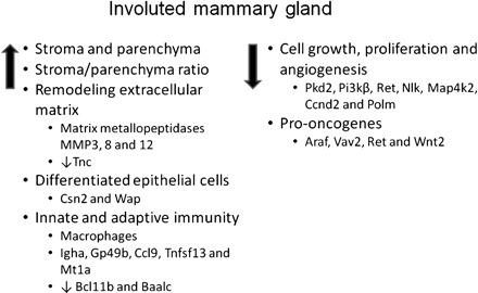
A full-term pregnancy early in life reduces lifetime risk of developing breast cancer, and the effect can be mimicked in rodents by full-term pregnancy or short-term treatment with exogenous estrogen and progesterone. To gain insight into the protective mechanism, 15 3-mo-old postpubertal virgin Lewis rats were randomly assigned to three groups: control (C), pregnancy (P), or hormone (H). The P group animals underwent a full-term pregnancy, and H group animals were implanted subcutaneously with silastic capsules filled with ethynyl estradiol and megesterol acetate for 21 days. C and P animals were implanted with sham capsules. On day 21 capsules were removed, which was followed by a 49-day involution period, euthanasia, and mammary tissue collection. Global gene expression was measured using Rat Genome 230.2 Arrays. Histological analysis revealed that P and H treatments induced sustained morphological changes in the mammary gland with significantly increased percentages of mammary parenchyma and stromal tissues and higher ratio of stroma to parenchyma. Transcriptome analysis showed that P and H treatments induced sustained global changes in gene expression in the mammary gland. Analysis of commonly up- and downregulated genes in P and H relative to C treatment showed increased expression of three matrix metallopeptidases (Mmp3, 8, and 12), more differentiated mammary phenotype, enhanced innate and adaptive immunity, and reduced cell proliferation and angiogenic signatures. The sustained morphological and global gene expression changes in mammary tissue after pregnancy and hormone treatment may function together to provide the protective effect against breast cancer.
Figures
Similar articles
-
Early exposure of the rat mammary gland to estrogen and progesterone blocks co-localization of estrogen receptor expression and proliferation.J Endocrinol. 2001 Oct;171(1):75-83. doi: 10.1677/joe.0.1710075. J Endocrinol. 2001. PMID: 11572792
-
Toward a physiological approach to breast cancer prevention.Cancer Epidemiol Biomarkers Prev. 1994 Jun;3(4):353-64. Cancer Epidemiol Biomarkers Prev. 1994. PMID: 8061586 Review.
-
Hormone-induced protection against mammary tumorigenesis is conserved in multiple rat strains and identifies a core gene expression signature induced by pregnancy.Cancer Res. 2006 Jun 15;66(12):6421-31. doi: 10.1158/0008-5472.CAN-05-4235. Cancer Res. 2006. PMID: 16778221
-
Hormonal prevention of breast cancer: mimicking the protective effect of pregnancy.Proc Natl Acad Sci U S A. 1999 Mar 2;96(5):2520-5. doi: 10.1073/pnas.96.5.2520. Proc Natl Acad Sci U S A. 1999. PMID: 10051675 Free PMC article.
-
A mouse mammary gland involution mRNA signature identifies biological pathways potentially associated with breast cancer metastasis.J Mammary Gland Biol Neoplasia. 2009 Jun;14(2):99-116. doi: 10.1007/s10911-009-9120-1. Epub 2009 Apr 30. J Mammary Gland Biol Neoplasia. 2009. PMID: 19408105 Review.
Cited by
-
Mimicking pregnancy as a strategy for breast cancer prevention.Breast Cancer Manag. 2013 Jul 1;2(4):283-294. doi: 10.2217/bmt.13.16. Breast Cancer Manag. 2013. PMID: 24738009 Free PMC article.
-
Association of breast cancer and obesity in a homogeneous population from Spain.J Endocrinol Invest. 2012 Jul;35(7):681-5. doi: 10.3275/8370. Epub 2012 Apr 18. J Endocrinol Invest. 2012. PMID: 22522745
-
The genomic signature of breast cancer prevention.Genes (Basel). 2014 Feb 26;5(1):65-83. doi: 10.3390/genes5010065. Genes (Basel). 2014. PMID: 24705287 Free PMC article.
-
Reproductive epidemiology of glial tumors may reveal novel treatments: high-dose progestins or progesterone antagonists as endocrino-immune modifiers against glioma.Neurosurg Rev. 2019 Jun;42(2):351-369. doi: 10.1007/s10143-018-0953-1. Epub 2018 Feb 17. Neurosurg Rev. 2019. PMID: 29453736 Review.
-
Reproductive history and breast cancer risk.Breast Cancer. 2012 Oct;19(4):302-8. doi: 10.1007/s12282-012-0384-8. Epub 2012 Jun 19. Breast Cancer. 2012. PMID: 22711317 Free PMC article. Review.
References
-
- Acuff HB, Sinnamon M, Fingleton B, Boone B, Levy SE, Chen X, Pozzi A, Carbone DP, Schwartz DR, Moin K, Sloane BF, Matrisian LM. Analysis of host- and tumor-derived proteinases using a custom dual species microarray reveals a protective role for stromal matrix metalloproteinase-12 in non-small cell lung cancer. Cancer Res 66: 7968– 7975, 2006 - PubMed
-
- American Cancer Society. Breast Cancer Facts & Figures 2009–2010, 2009
-
- Balbin M, Fueyo A, Tester AM, Pendas AM, Pitiot AS, Astudillo A, Overall CM, Shapiro SD, Lopez-Otin C. Loss of collagenase-2 confers increased skin tumor susceptibility to male mice. Nat Genet 35: 252– 257, 2003 - PubMed
-
- Baldus CD, Tanner SM, Kusewitt DF, Liyanarachchi S, Choi C, Caligiuri MA, Bloomfield CD, de la Chapelle A. BAALC, a novel marker of human hematopoietic progenitor cells. Exp Hematol 31: 1051– 1056, 2003 - PubMed
Publication types
MeSH terms
Grants and funding
LinkOut - more resources
Full Text Sources
Medical
Molecular Biology Databases
Miscellaneous