. 2011 Nov 15;25(22):2361-73.
doi: 10.1101/gad.174110.111.

Maelstrom coordinates microtubule organization during Drosophila oogenesis through interaction with components of the MTOC

Affiliations

Maelstrom coordinates microtubule organization during Drosophila oogenesis through interaction with components of the MTOC

Kaoru Sato et al. Genes Dev. .

Abstract

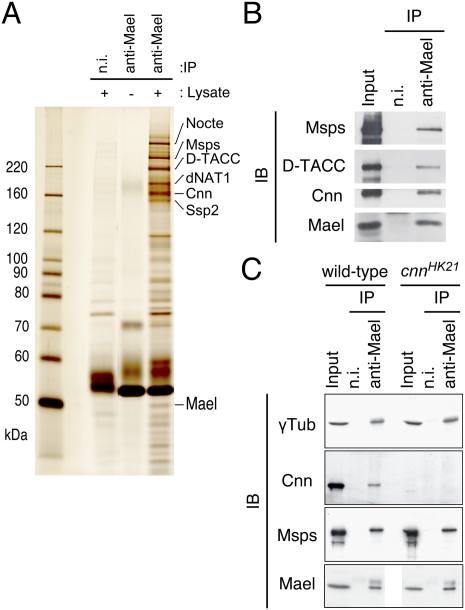
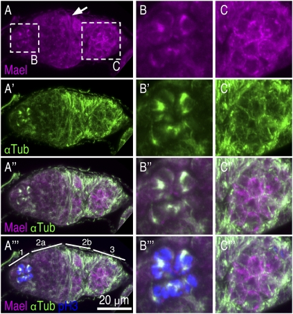
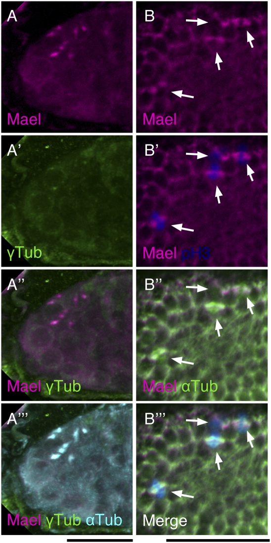
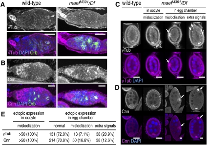
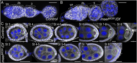
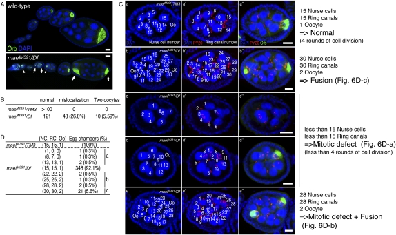
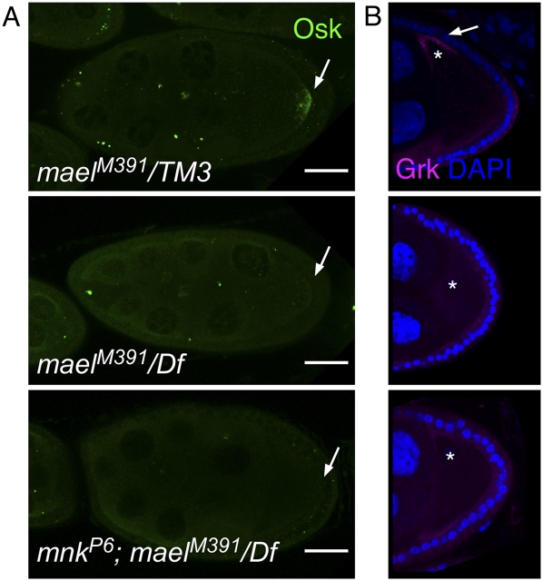
The establishment of body axes in multicellular organisms requires accurate control of microtubule polarization. Mutations in Drosophila PIWI-interacting RNA (piRNA) pathway genes often disrupt the axes of the oocyte. This results from the activation of the DNA damage checkpoint factor Checkpoint kinase 2 (Chk2) due to transposon derepression. A piRNA pathway gene, maelstrom (mael), is critical for the establishment of oocyte polarity in the developing egg chamber during Drosophila oogenesis. We show that Mael forms complexes with microtubule-organizing center (MTOC) components, including Centrosomin, Mini spindles, and γTubulin. We also show that Mael colocalizes with αTubulin and γTubulin to centrosomes in dividing cyst cells and follicle cells. MTOC components mislocalize in mael mutant germarium and egg chambers, leading to centrosome migration defects. During oogenesis, the loss of mael affects oocyte determination and induces egg chamber fusion. Finally, we show that the axis specification defects in mael mutants are not suppressed by a mutation in mnk, which encodes a Chk2 homolog. These findings suggest a model in which Mael serves as a platform that nucleates other MTOC components to form a functional MTOC in early oocyte development, which is independent of Chk2 activation and DNA damage signaling.


Figures

Figure 1.
Figure 1.
Mael forms complexes with MTOC components in ovaries. (A) Immunoprecipitation was performed from ovary lysates using an anti-Mael monoclonal antibody. Proteins immunoprecipitated were stained with silver. Mass spectrometry analyses revealed that Nocte, Msps, D-TACC, dNAT1, Cnn, and Ssp2 coimmunoprecipitated with Mael. “n.i.” indicates nonimmune IgG used as a negative control. (B) Western blotting on the Mael immunoprecipitated complex in A using anti-Msps, anti-D-TACC, and anti-Cnn confirmed Mael binding with Msps, D-TACC, and Cnn. Nonimmune IgG (n.i.) was used as a negative control. (C) Immunoprecipitation was performed from wild-type and cnn mutant ovary lysates using an anti-Mael monoclonal antibody. cnnHK21 is a cnn-null allele with a nonsense mutation that truncates the protein at amino acid 106 (Megraw et al. 1999). Mael binds with γTub and Msps in a Cnn-independent manner.
Figure 2.
Figure 2.
Mael colocalizes with αTub in the germarium. (A–A″′) Ovaries were immunostained with anti-Mael, anti-αTub, and anti-pH3 antibodies. pH3 is a mitotic marker. In region 1 of the germarium, Mael accumulates in discrete regions of dividing cyst cells. The Mael signals overlaps with the γTub signals, suggesting that Mael is localized to the MT spindles in early cyst cells. In region 2, Mael is localized to granules, in which γTub was undetected. In region 3, Mael is accumulated in the nuage, which is also devoid of γTub. A Mael signal in a somatic precursor cell is shown by an arrow. (B–B″′) Magnified views of dividing cyst cells in A–A′″′. (C–C″′) Magnified views of the early egg chamber in A–A″′.
Figure 3.
Figure 3.
Mael is localized to mitotic spindles in the dividing cyst and follicle cells. (A–A″′) Immunofluorescence was performed using anti-Mael, anti-αTub, and anti-γTub antibodies. Mael is colocalized with αTub and γTub at mitotic spindles of dividing cyst cells. (B–B″′) Immunofluorescence was performed using anti-Mael, anti-αTub, and anti-pH3 antibodies. In dividing follicle cells (pH3-positive cells; indicated by arrows), Mael is colocalized with αTub to mitotic spindles. Bars, 20 μm.
Figure 4.
Figure 4.
Protein components of the MTOC are mislocalized in mael mutant ovaries. (A) γTub localization in the germarial region of wild-type (left panels) and mael mutant (right panels) ovaries. Anti-Orb antibody was used as a marker for developing oocytes. γTub is accumulated in the oocyte in the wild-type germarium (arrow), but was absent from the mael mutant oocyte in the germarium. (B) Cnn localization in the germarial region of wild-type (left panels) and mael mutant ovaries (right panels). Cnn, which is undetectable in the oocyte in the wild-type germarium, was detected in the oocytes in the mael mutant germarium (arrows). (C) γTub localization in wild-type and mael mutant egg chambers. In the wild type, γTub is specifically accumulated at the anterior region in the oocyte. However, in the mael mutant, γTub was often mislocalized to the posterior region of the oocytes. γTub was also ectopically accumulated within the egg chambers (arrows) of mael. (D) Cnn localization in wild-type and mael mutant egg chambers. Cnn, which is accumulated at the posterior region in the wild-type oocyte, was detected as speckles in the mael mutant oocytes. Cnn was ectopically accumulated within the egg chambers (arrows). (E) The percentage of mael egg chambers that show ectopic localization of Cnn and γTub. Bars, 20 μm.
Figure 5.
Figure 5.
MTOC formation is defective in mael mutant ovaries. MT distribution in wild-type and mael mutant (maelM391/Df) oocytes was visualized with anti-αTub antibody (white). DNA was visualized with DAPI (blue). (A) MTs in the control germarium and stage 1 egg chamber. The MTOC was observed at the posterior region of the oocyte in region 3 of the germarium. A red asterisk indicates the nucleus of the oocyte that is denoted by a dotted line. (B) MTs in the mael mutant germarium and stage 1 egg chamber. The MTOC was not detected when mael function was lost. (C) MTs in stage 3–8 egg chambers in control and mael mutant ovaries. The MTOC was detected at the posterior region of stage 3–6 control oocytes, but not in the mael mutant oocytes. In the wild type, MTs are then rolled up (“diaphragm” pattern) during stage 7 (Supplemental Fig. S1) and project from the oocyte nucleus in an anterior–posterior direction at stage 8 (“open diaphragm” pattern). This reorganization of MTs was not observed in the mael mutant. Bars, 20 μm.
Figure 6.
Figure 6.
Defective egg chamber development in the mael mutant. (A) Wild-type (top) and mael mutant (bottom) oocytes were visualized by immunohistochemical analyses using an anti-Orb antibody (green). DNA was stained with DAPI (blue). In the wild type, the oocyte is localized at the posterior region of the egg chamber. In contrast, in the mael mutant, egg chambers possessing a mislocalized oocyte (arrowheads) or two oocytes (arrows) were observed. (B) The percentage of abnormal egg chambers in the mael mutant. (C) The number of nurse cells and ring canals in the mael mutant egg chambers. Ring canals (red) and oocytes (green) were visualized using PY20 and anti-Orb antibody, respectively. DNA was stained with DAPI (blue). (D) The population of abnormal egg chambers in the mael mutants. NC, RC, and Oo indicate the number of nurse cells, ring canals, and oocytes, respectively. (Groups a–c) See the text. Bars, 20 μm.
Figure 7.
Figure 7.
mnk does not suppress the mael phenotype. (A) Mutations in mnk do not suppress the Osk localization defect in mael mutant ovaries. (Top) In the control oocyte (stage 9), Osk is localized at the posterior region of the oocyte (arrow). (Middle) In mael egg chambers, the localization of Osk was dispersed throughout the oocyte. (Bottom) In mnk;mael double mutant egg chambers, the mislocalization of Osk was not rescued. (B) In the control, Grk is localized at the dorsal anterior cortex (arrow) near the oocyte nucleus (asterisk). (Bottom) In mnk;mael double mutant egg chambers, the localizations of Osk and Grk are also dispersed throughout the oocyte. (Middle) In mael egg chambers, Grk was not accumulated. (Bottom) In mnk;mael double mutant egg chambers, the mislocalization of Grk was not rescued. DAPI (blue) marks the cell nuclei. Bars, 20 μm.

References

    1. Adams RR, Maiato H, Earnshaw WC, Carmena M 2001. Essential roles of Drosophila inner centromere protein (INCENP) and aurora B in histone H3 phosphorylation, metaphase chromosome alignment, kinetochore disjunction, and chromosome segregation. J Cell Biol 153: 865–880 - PMC - PubMed
    1. Aravin AA, Naumova NM, Tulin AV, Vagin VV, Rozovsky YM, Gvozdev VA 2001. Double-stranded RNA-mediated silencing of genomic tandem repeats and transposable elements in the D. melanogaster germline. Curr Biol 11: 1017–1027 - PubMed
    1. Arn EA, Cha BJ, Theurkauf WE, Macdonald PM 2003. Recognition of a bicoid mRNA localization signal by a protein complex containing Swallow, Nod, and RNA binding proteins. Dev Cell 4: 41–51 - PubMed
    1. Bolívar J, Huynh JR, López-Schier H, González C, St Johnston D, González-Reyes A 2001. Centrosome migration into the Drosophila oocyte is independent of BicD and egl, and of the organisation of the microtubule cytoskeleton. Development 128: 1889–1897 - PubMed
    1. Brendza RP, Serbus LR, Duffy JB, Saxton WM 2000. A function for kinesin I in the posterior transport of oskar mRNA and Staufen protein. Science 289: 2120–2122 - PMC - PubMed

Publication types

MeSH terms

LinkOut - more resources