. 2011 Nov 15;25(22):2374-86.
doi: 10.1101/gad.178079.111.

Drosophila CLOCK target gene characterization: implications for circadian tissue-specific gene expression

Affiliations

Drosophila CLOCK target gene characterization: implications for circadian tissue-specific gene expression

Katharine Compton Abruzzi et al. Genes Dev. .

Abstract

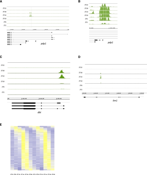
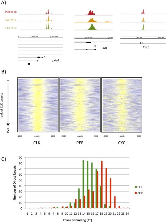
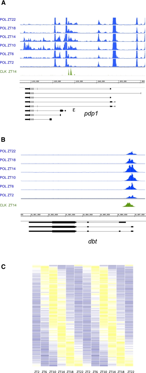
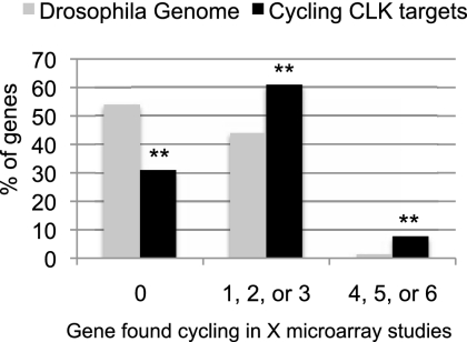
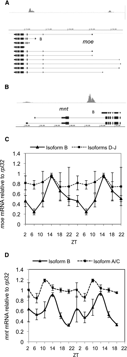
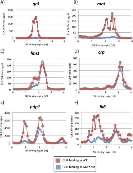
CLOCK (CLK) is a master transcriptional regulator of the circadian clock in Drosophila. To identify CLK direct target genes and address circadian transcriptional regulation in Drosophila, we performed chromatin immunoprecipitation (ChIP) tiling array assays (ChIP-chip) with a number of circadian proteins. CLK binding cycles on at least 800 sites with maximal binding in the early night. The CLK partner protein CYCLE (CYC) is on most of these sites. The CLK/CYC heterodimer is joined 4-6 h later by the transcriptional repressor PERIOD (PER), indicating that the majority of CLK targets are regulated similarly to core circadian genes. About 30% of target genes also show cycling RNA polymerase II (Pol II) binding. Many of these generate cycling RNAs despite not being documented in prior RNA cycling studies. This is due in part to different RNA isoforms and to fly head tissue heterogeneity. CLK has specific targets in different tissues, implying that important CLK partner proteins and/or mechanisms contribute to gene-specific and tissue-specific regulation.


Figures

Figure 1.
Figure 1.
CLK binding to direct target genes peaks at ZT14. CLK ChIPs were performed at six different time points throughout the day, and the resulting DNA was analyzed using tiling arrays (Affymetrix). CLK binding is visualized using the IGB. Genes above the genomic coordinates are transcribed from left to right (plus strand), and genes below the genomic coordinates are transcribed from right to left (minus strand). CLK binds rhythmically to the promoters of pdp1 (primarily the ε isoform) (A), of pdp1 (primarily the ε isoform) zoomed in to show binding even at ZT2 (B), of dbt (C), and in the middle of lim1 (D). CLK binding peaks at ZT14 on these three genes. (E) CLK binding cycles on ∼800 genes. Genes were sorted by binding phase, and CLK ChIP signal is portrayed using a heat map in which data for a 24-h period are concatenated to show cycling. CLK ChIP signal ranges from low (dark blue; Z-score between −2 and −0.5; i.e., between 2 and 0.5 standard deviations below the mean) to medium (white; Z-score between −0.5 and +0.5) to highest (yellow; Z-score between 0.5 and 2). For most genes, the highest CLK ChIP signal is at ZT14.
Figure 2.
Figure 2.
CLK direct targets also bind PER and CYC. (A) CLK (green; ZT14), CYC (orange; ZT14), and PER (red; ZT18) are all bound in nearly identical locations on pdp1ε, dbt, and lim1. (B) The ChIP signals of CLK, PER, and CYC in regions of CLK binding are represented using a heat map. As expected, CLK ChIP signals are strong (yellow; Z-score between 2 and 6) in the middle of the CLK peaks. The PER ChIP signal is also high (yellow; Z-score between 2 and 6) in these regions, suggesting that PER binds with CLK on the majority of genes. Despite a lower CYC ChIP strength, CYC binding is also enriched where CLK binds (yellow; Z-score between 1.5 and 6). This suggests that the majority of CLK direct targets are also bound by PER and CYC. (C) Histogram showing the phase of cycling CLK (green) and PER (red) binding as determined by Fourier analysis (see the Materials and Methods). CLK binding precedes PER binding by ∼4–6 h.
Figure 3.
Figure 3.
Approximately 30% of CLK direct target genes have cycling Pol II on their promoters and/or in their coding regions. Pol II ChIP–chips were performed on fly head chromatin collected every 4 h for a total of six time points. (A,B) Pol II binding is visualized on the IGB (Affymetrix). Genes above the genomic coordinates are transcribed from left to right (plus strand), and genes below the genomic coordinates are transcribed from right to left (minus strand). For comparison, CLK binding is shown in green. (A) Pol II occupancy on pdp1 shows both cycling promoter-proximal Pol II binding and cyclical binding of Pol II in the coding region of the circadian-controlled ε isoform. (B) Cyclical promoter-proximal Pol II binding is observed on the circadian kinase dbt. Pol II is always present at the promoter but increases to peak between ZT10 and ZT14. No cyclical Pol II in the coding region is detectable. (C) Heat map showing the Pol II ChIP signal across circadian time on those genes that have overlapping Pol II and CLK peaks (<30% of cycling CLK peaks). Data are double-plotted to aid in the visualization of cycling. Lowest ChIP signal is shown in blue (Z-score −6 to −2), and highest ChIP signal is shown in yellow (Z-score 2–6). Most Pol II peaks oscillate with a phase of ZT10–ZT14. Very few Pol II peaks are maximal at ZT22 or ZT0.
Figure 4.
Figure 4.
CLK direct targets are enriched for cycling genes. Six different circadian microarray data sets were analyzed and cycling genes were identified. We categorized genes as cycling (identified in four, five, or six independent studies), inconsistently cycling (identified in one, two, or three independent studies) or not cycling (never identified in a study). The graph shows the distribution of all the of genes in the Drosophila genome (gray) or CLK direct target genes (black; only those with cycling binding) into these categories. CLK direct targets are enriched in cycling genes (identified in four or more studies) as well as genes inconsistently identified to cycle (identified in one, two, or three studies). They also show a substantial decrease in the number of genes not identified as cycling in any of the six studies. Double asterisks indicate that the difference between CLK direct targets and the genome as a whole is statistically significant (P-value <10−4).
Figure 5.
Figure 5.
CLK binding results in isoform-specific mRNA cycling. Fifty-five percent of CLK direct targets have alternative start sites. (A) CLK binds to both the short (isoform B) and long isoforms of moe at ZT14. (B) CLK binds to the short isoform (isoform B) of mnt at ZT14. (C) mRNA levels at six time points throughout the day are double-plotted and show that the mRNA resulting from isoform B of moe cycles with a peak amplitude at ZT14 (triangles; solid line). In contrast, mRNAs resulting from the longer isoforms (isoforms D–J) of moe do not cycle (squares; dashed line). (D) mRNA levels at six time points throughout the day are double-plotted and show that the mRNA resulting from isoform B of mnt cycles with a peak amplitude at ZT14 (triangles; solid line). In contrast, mRNAs resulting from the longer isoforms (isoforms A and C) of mnt do not cycle (squares; dashed line).
Figure 6.
Figure 6.
CLK binding can be tissue-specific. CLK ChIP–chips were performed on either wild-type (CLK-V5) or GMR-hid (CLK-V5, GMR-hid) flies at ZT14. In GMR-hid flies, the majority of the eye tissue is ablated. We compare CLK ChIP signal in wild-type (red) and GMR-hid (blue) flies on six different genes. CLK binding is undetectable on gol (A) and mnt (B) when eyes are ablated, suggesting that gol and mnt are CLK direct targets primarily in eye tissue. CLK binding is unaffected on lim1 (C) and crp (D) in GMR-hid, suggesting that these genes are CLK targets in non-eye tissue. Interestingly, on some genes with multiple CLK promoter peaks, the two peaks of CLK binding are differentially affected in GMR-hid. One of the peaks of CLK binding on both pdp1 (E) and lk6 (F) is greatly diminished in GMR-hid, while another peak is much less affected.

Comment in

Similar articles

Cited by

References

    1. Allada R, White N, So W, Hall J, Rosbash M 1998. A mutant Drosophila homolog of mammalian Clock disrupts circadian rhythms and transcription of period and timeless. Cell 93: 791–804 - PubMed
    1. Antoch MP, Song E-J, Chang A-M, Vitaterna MH, Zhao Y, Wilsbacher LD, Sangoram AM, King DP, Pinto LH, Takahashi JS 1997. Functional identification of the mouse circadian clock gene by transgenic BAC rescue. Cell 89: 655–667 - PMC - PubMed
    1. Blau J, Young MW 1999. Cycling vrille expression is requiered for a functional Drosophila clock. Cell 99: 661–671 - PubMed
    1. Chiu JC, Ko HW, Edery I 2011. NEMO/NLK phosphorylates PERIOD to initiate a time-delay phosphorylation circuit that sets circadian clock speed. Cell 145: 357–370 - PMC - PubMed
    1. Curtin K, Huang ZJ, Rosbash M 1995. Temporally regulated nuclear entry of the Drosophila period protein contributes to the circadian clock. Neuron 14: 365–372 - PubMed

Publication types

MeSH terms