. 2012 Jan 1;52(1):70-8.
doi: 10.1016/j.visres.2011.10.016. Epub 2011 Nov 9.

Isoeccentric locations are not equivalent: the extent of the vertical meridian asymmetry

Affiliations

Isoeccentric locations are not equivalent: the extent of the vertical meridian asymmetry

Jared Abrams et al. Vision Res. .

Abstract

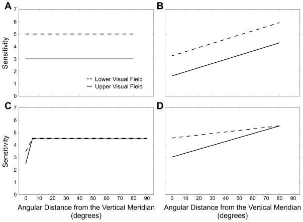
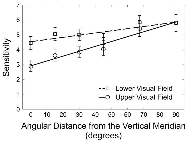
Performance in visual tasks is limited by the low-level mechanisms that sample the visual field. It is well documented that contrast sensitivity and spatial resolution decrease as a function of eccentricity and that those factors impair performance in "higher level" tasks, such as visual search. Performance also varies consistently at isoeccentric locations in the visual field. Specifically, at a fixed eccentricity, performance is better along the horizontal meridian than the vertical meridian, and along the lower than the upper vertical meridian. Whether these asymmetries in visual performance fields are confined to the vertical meridian or extend across the whole upper versus lower visual hemifield has been a matter of debate. Here, we measure the extent of the upper versus lower asymmetry. Results reveal that this asymmetry is most pronounced at the vertical meridian and that it decreases gradually as the angular distance (polar angle) from the vertical meridian increases, with eccentricity held constant. Beyond 30° of polar angle from the vertical meridian, the upper to lower asymmetry is no longer reliable. Thus, the vertical meridian is uniquely asymmetric and uniquely insensitive. This pattern of results is consistent with early anatomical properties of the visual system and reflects constraints that are critical to our understanding of visual information processing.


Figures

Fig. 1
Fig. 1
Four possible patterns for upper and lower visual field sensitivity as a function of angular distance from the vertical meridian. Sensitivity is the reciprocal of the contrast necessary to reach 75% performance in the orientation discrimination task. (A) One possibility is a constant upper to lower asymmetry where the vertical meridian plays no role. (B) Alternatively, there may be a constant upper to lower asymmetry where performance is worst at the vertical meridian. (C) Conversely, it is possible that there is no upper versus lower asymmetry, with an upper to lower difference that is restricted to the vertical meridian. (D) Finally, it may be that the vertical meridian is the most asymmetric and insensitive portion of the visual field and that the asymmetry decreases as a function of angular distance from the vertical meridian.
Fig. 2
Fig. 2
Trial sequence. Observers performed a 2AFC orientation discrimination on Gabor stimuli. Note that only four locations were present on a given trial and that only four of the possible locations are represented by this figure. Note also that the Gabor tilt angle, as well as the size of the place-holders, fixation point, and response cue have been exaggerated for clarity.
Fig. 3
Fig. 3
(a) HVA and VMA across observers. The HVA as the raw difference between averaged sensitivity (1/threshold) on the horizontal meridian and the vertical meridian and the VMA as the difference between sensitivity (1/threshold) along the lower and the upper vertical meridian. Values greater than zero indicate higher performance along the horizontal meridian and higher performance along the lower than the upper vertical meridian, respectively. Error bars represent 95% confidence intervals of the difference. (b) HVA for individual observers. The HVA for each observer is depicted as the sensitivity along the horizontal meridian as a function of sensitivity along the vertical meridian. Points above the diagonal indicate that sensitivity is lower along the vertical meridian than the horizontal meridian. (c) VMA for individual observers. The VMA for each observer is depicted as the sensitivity along the lower vertical meridian as a function of sensitivity along the upper vertical meridian. Points above the diagonal indicate that sensitivity is lower along the upper vertical meridian than the lower vertical meridian.
Fig. 4
Fig. 4
Sensitivity (1/threshold) as a function of angular distance from the vertical meridian for the upper and lower visual hemifields, as well as a fit to both sets of data using linear regression. Error bars represent ±1 standard error of the mean.

References

    1. Adams DL, Horton JC. A precise retinotopic map of primate striate cortex generated from the representation of angioscotomas. Journal of Neuroscience. 2003;23(9):3771–3789. - PMC - PubMed
    1. Altpeter E, Mackeben M, Trauzettel-Klosinski S. The importance of sustained attention for patients with maculopathies. Vision Research. 2000;40(10–12):1539–1547. - PubMed
    1. Anderson RS, Wilkinson MO, Thibos LN. Psychophysical localization of the human visual streak. Optometry and Vision Science. 1992;69(3):171–174. - PubMed
    1. Bouma H. Interaction effects in parafoveal letter recognition. Nature. 1970;226(5241):177–178. - PubMed
    1. Brainard DH. The psychophysics toolbox. Spatial Vision. 1997;10(4):433–436. - PubMed

Publication types