. 2012 Feb;13(1):1-16.
doi: 10.1007/s10162-011-0297-9. Epub 2011 Nov 16.

Brain-derived neurotrophic factor modulates auditory function in the hearing cochlea

Affiliations

Brain-derived neurotrophic factor modulates auditory function in the hearing cochlea

David J Sly et al. J Assoc Res Otolaryngol. 2012 Feb.

Abstract

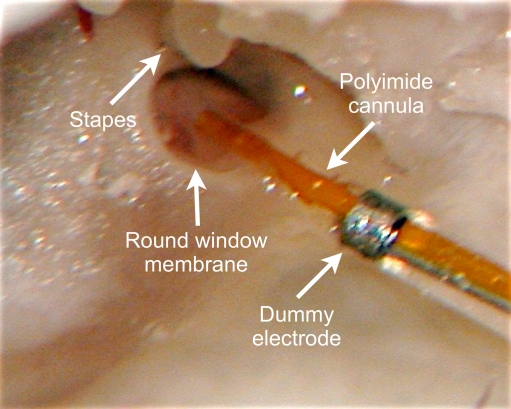
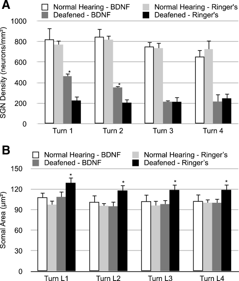
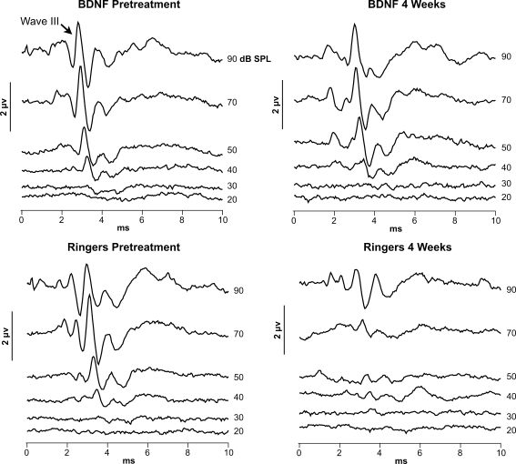
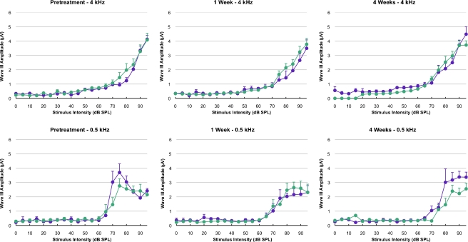
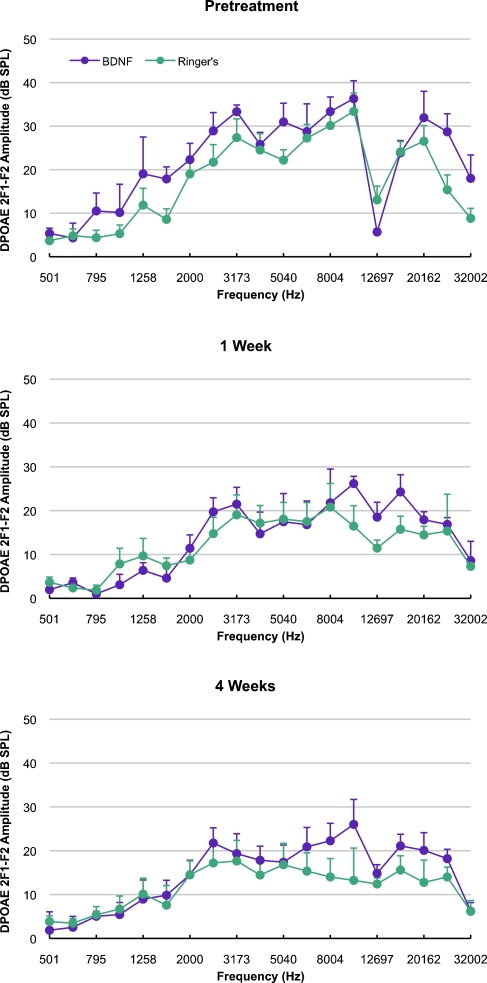
Neurotrophins prevent spiral ganglion neuron (SGN) degeneration in animal models of ototoxin-induced deafness and may be used in the future to improve the hearing of cochlear implant patients. It is increasingly common for patients with residual hearing to undergo cochlear implantation. However, the effect of neurotrophin treatment on acoustic hearing is not known. In this study, brain-derived neurotrophic factor (BDNF) was applied to the round window membrane of adult guinea pigs for 4 weeks using a cannula attached to a mini-osmotic pump. SGN survival was first assessed in ototoxically deafened guinea pigs to establish that the delivery method was effective. Increased survival of SGNs was observed in the basal and middle cochlear turns of deafened guinea pigs treated with BDNF, confirming that delivery to the cochlea was successful. The effects of BDNF treatment in animals with normal hearing were then assessed using distortion product otoacoustic emissions (DPOAEs), pure tone, and click-evoked auditory brainstem responses (ABRs). DPOAE assessment indicated a mild deficit of 5 dB SPL in treated and control groups at 1 and 4 weeks after cannula placement. In contrast, ABR evaluation showed that BDNF lowered thresholds at specific frequencies (8 and 16 kHz) after 1 and 4 weeks posttreatment when compared to the control cohort receiving Ringer's solution. Longer treatment for 4 weeks not only widened the range of frequencies ameliorated from 2 to 32 kHz but also lowered the threshold by at least 28 dB SPL at frequencies ≥16 kHz. BDNF treatment for 4 weeks also increased the amplitude of the ABR response when compared to either the control cohort or prior to treatment. We show that BDNF applied to the round window reduces auditory thresholds and could potentially be used clinically to protect residual hearing following cochlear implantation.


Figures

FIG. 1
FIG. 1
Drug delivery to the round window membrane. A polyimide cannula connected to PVC tubing was placed near the intact round window membrane to deliver chronic drug treatments to the cochlea. A dummy electrode was used to guide the depth of insertion of the cannula into the bulla. The cannula was connected to a mini-osmotic pump (not shown) located under the skin at the scapulae and the pump delivered treatments over 4 weeks.
FIG. 2
FIG. 2
Representative photomicrographs of the lower basal turn of the cochlea. Each image shows a representative section from one animal in each of the four experimental groups. Each inset shows higher-magnification images of the organ of Corti. In the deafened animals, the organ of Corti was degenerated and there were typically no remaining hair cells, while normal hearing animals treated with BDNF or Ringer’s solution had intact inner and outer hair cells. In deafened animals, those treated with BDNF had greater preservation of peripheral dendrites in the osseous spiral lamina and SGNs in Rosenthal’s canal, evidenced in the images by less white space in these regions. Normal hearing animals had the densest packing of neural tissue within the osseous spiral lamina and Rosenthal’s canal, with no differences evident between those animals treated with either BDNF or Ringer’s solution.
FIG. 3
FIG. 3
Representative photomicrographs of SGNs in lower basal turn of the cochlea. These photomicrographs are higher-magnification images of those shown in Figure 2. The region of the Rosenthal’s canal containing SGNs is outlined in the lower right section. In deafened animals, the density of SGNs was greater in animals treated with BDNF than those treated with Ringer’s solution. In normal hearing animals, the density of SGNs was greater than that of the deafened animals and the SGNs showed less degeneration and more compact nuclei. The density of SGNs among normal hearing animals was similar in those treated with either BDNF or Ringer’s solution.
FIG. 4
FIG. 4
The effect of treatment on SGN A density and B somal area. Each bar represents the mean ± SEM density averaged across all animals in each group. In Aasterisks represent groups significantly (p < 0.01) different from deafened Ringer’s control group and in Basterisks represent groups significantly (p < 0.05) greater than all other groups. Five sections per animal were used for SGN density and between six and nine sections per animal were used for somal area estimations. Only the lower portions of cochlear turns were used for somal area estimations. The somal area sample was a relatively large sample of 7,470 soma across 24 animals. Deafened guinea pigs treated with BDNF through the round window membrane had a significantly greater preservation of SGNs in the lower turns of the cochlear spiral (numbered 1 and 2, turn 1 being the most basal) compared to deafened animals treated with Ringer’s solution, suggesting that BDNF was successfully delivered across the intact round window membrane to the basal regions of the cochlea. Normal hearing guinea pigs treated with BDNF had similar SGN densities to normal hearing animals treated with Ringer’s solution; the SGN density of each of these groups was considerably larger in turns 1 and 2 than the SGN density of deafened animals treated with BDNF. Deafened guinea pigs left treated with Ringer’s solution had larger somal areas in all turns compared to all other groups. Normal hearing animals treated with BDNF had larger somal areas compared to normal hearing animals treated with Ringer’s solution in turns 1–3, with the greatest difference occurring in turn 1.
FIG. 5
FIG. 5
Example ABR responses to acoustic click stimulation in normal hearing animals. Prior to treatment, the amplitude of wave III of the ABR response was similar in animals within the BDNF or Ringer’s group. By 4 weeks posttreatment, the amplitude and threshold of wave III had reduced in animals treated with Ringer’s solution, but in BDNF-treated animals the response remained similar to that prior to treatment. While there was a characteristic reduction in the latency (time of occurrence) of wave III with increasing stimulus amplitude, there was no difference in the latency of this wave among the groups.
FIG. 6
FIG. 6
The effect of treatment on ABR threshold. Each data point represents the mean ± SEM threshold across animals. Note that the data for clicks are presented as individual points on the right side of each graph. Prior to implant, there was little difference between treatments, with both BDNF- and Ringer’s-treated animals having the lowest thresholds between 4 and 16 kHz. Immediately post-surgery, the thresholds in both groups had risen by 5–10 dB SPL across most stimulus frequencies, including clicks. After 1 week, the thresholds of both groups had dropped, although those for BDNF had dropped more. After 4 weeks, the threshold in the Ringer’s group had stabilized, being similar to those at 1 week, but not fully recovering to pretreatment levels in the mid-high frequencies (4–16 kHz). However, after 4 weeks, the thresholds for the BDNF group dropped further to the point where they were below pretreatment levels and up to 29 dB SPL lower than the Ringer’s group in the mid-high frequencies (2–32 kHz). Note that the data recorded immediately post-surgery were not included in statistical analyses as sufficient data were not recorded in all animals.
FIG. 7
FIG. 7
The effect of treatment on ABR amplitude. Each data point represents the mean ± SEM of the amplitude of wave III of the auditory brainstem response. Prior to implant, there were no differences in the amplitude growth function between the BDNF and Ringer’s treatment groups. The rising function of the ABR amplitude with increasing stimulus intensity was similar across all the pure tone frequencies; the response to clicks starts at lower intensities and rises to greater amplitudes, reflecting the broader excitation of the click stimulus across the cochlea (note the different vertical axis scales for the click and tone pip data). After 4 weeks of treatment, the ABR amplitudes in BDNF-treated animals exceeded pretreatment levels. However, over the same period, the ABR amplitudes in the Ringer’s group had dropped across most frequencies.
FIG. 7
FIG. 7
The effect of treatment on ABR amplitude. Each data point represents the mean ± SEM of the amplitude of wave III of the auditory brainstem response. Prior to implant, there were no differences in the amplitude growth function between the BDNF and Ringer’s treatment groups. The rising function of the ABR amplitude with increasing stimulus intensity was similar across all the pure tone frequencies; the response to clicks starts at lower intensities and rises to greater amplitudes, reflecting the broader excitation of the click stimulus across the cochlea (note the different vertical axis scales for the click and tone pip data). After 4 weeks of treatment, the ABR amplitudes in BDNF-treated animals exceeded pretreatment levels. However, over the same period, the ABR amplitudes in the Ringer’s group had dropped across most frequencies.
FIG. 8
FIG. 8
The effect of treatment on DPOAE amplitude. Each point represents the mean ± SEM DPOAE amplitude in response to a 65-dB SPL stimulus. The amplitude is the 2F1–F2 distortion product, which was typically the largest distortion product response. The frequency is the geometric mean of F1 and F2 stimulus frequencies. Prior to treatment, the DPOAE response amplitudes were slightly larger in the BDNF group. No significant differences were found between treatments. There was a drop in DPOAE amplitude across most frequencies in both groups by 1 week that stabilized by 4 weeks. The diminished responses seen at 12 and 32 kHz in most of the data reflect our observations that DPOAEs were regularly lower at these frequencies in each guinea pig.

References

    1. Adamson CL, Reid MA, Davis RL. Opposite actions of brain-derived neurotrophic factor and neurotrophin-3 on firing features and ion channel composition of murine spiral ganglion neurons. J Neurosci. 2002;22:1385–1396. - PMC - PubMed
    1. Agterberg MJ, Versnel H, Groot JC, Smoorenburg GF, Albers FW, Klis SF. Morphological changes in spiral ganglion cells after intracochlear application of brain-derived neurotrophic factor in deafened guinea pigs. Hear Res. 2008;244:25–34. doi: 10.1016/j.heares.2008.07.004. - DOI - PubMed
    1. Coggeshall RE, Lekan HA. Methods for determining numbers of cells and synapses: a case for more uniform standards of review. J Comp Neurol. 1996;364:6–15. doi: 10.1002/(SICI)1096-9861(19960101)364:1<6::AID-CNE2>3.0.CO;2-9. - DOI - PubMed
    1. Davis-Lopez de Carrizosa MA, Morado-Diaz CJ, Tena JJ, Benitez-Temino B, Pecero ML, Morcuende SR, Cruz RR, Pastor AM. Complementary actions of BDNF and neurotrophin-3 on the firing patterns and synaptic composition of motoneurons. J Neurosci. 2009;29:575–587. doi: 10.1523/JNEUROSCI.5312-08.2009. - DOI - PMC - PubMed
    1. Diao MF, Gao WY, Sun JJ, Liu Y, Chen DL, Jiang W, Zhao J, Chen X. Protection from noise-induced hearing loss by a nitric oxide synthase inhibitor and neurotrophin 3 in the guinea pig cochlea. Zhonghua Er Bi Yan Hou Tou Jing Wai Ke Za Zhi. 2007;42:281–285. - PubMed

Publication types

MeSH terms

Substances