Comparative Study
. 2012 Jan 3;125(1):100-12.
doi: 10.1161/CIRCULATIONAHA.111.042598. Epub 2011 Nov 15.

Safety and efficacy of allogeneic cell therapy in infarcted rats transplanted with mismatched cardiosphere-derived cells

Affiliations
Comparative Study

Safety and efficacy of allogeneic cell therapy in infarcted rats transplanted with mismatched cardiosphere-derived cells

Konstantinos Malliaras et al. Circulation. .

Abstract

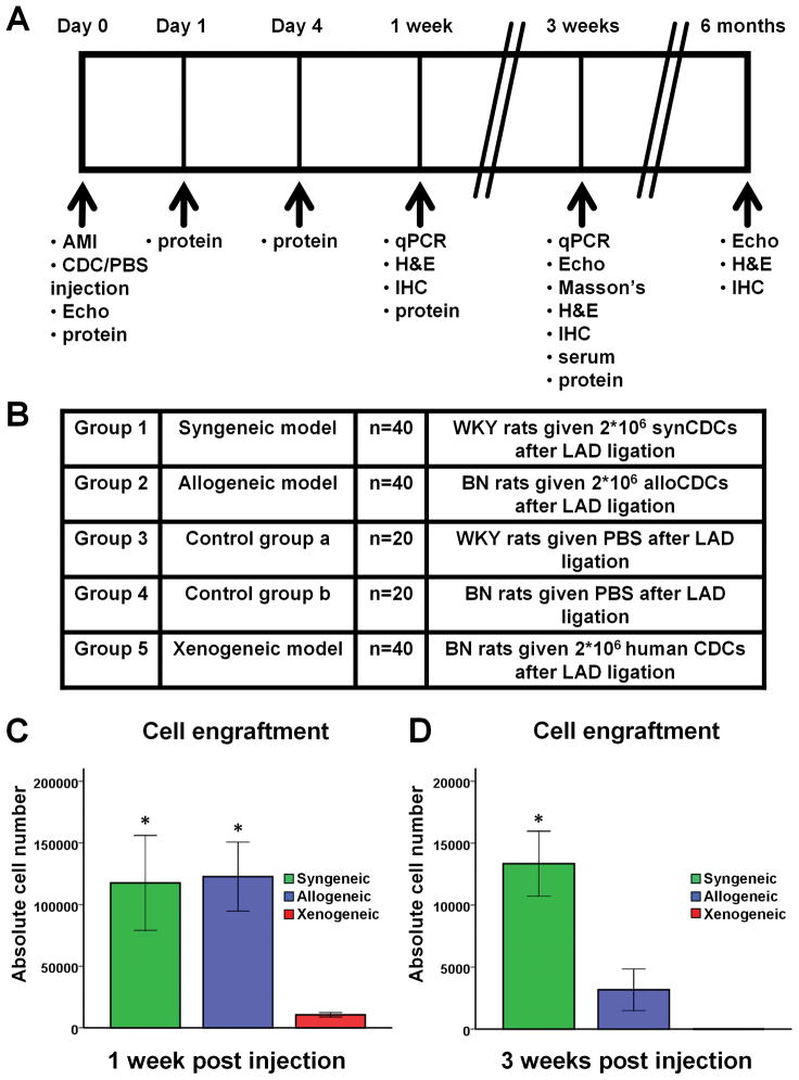
Background: Cardiosphere-derived cells (CDCs) are an attractive cell type for tissue regeneration, and autologous CDCs are being tested clinically. However, autologous therapy necessitates patient-specific tissue harvesting and cell processing, with delays to therapy and possible variations in cell potency. The use of allogeneic CDCs, if safe and effective, would obviate such limitations. We compared syngeneic and allogeneic CDC transplantation in rats from immunologically-mismatched inbred strains.

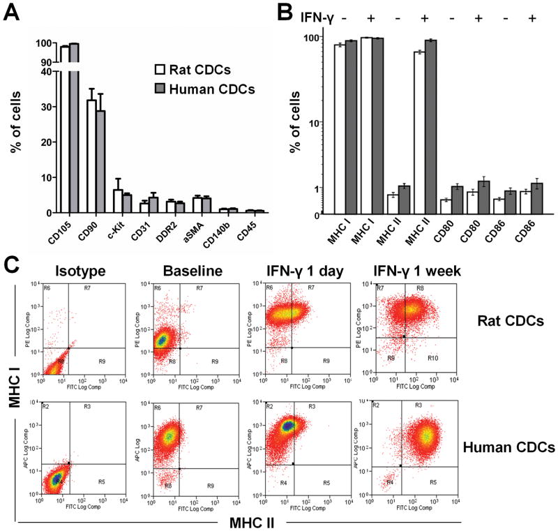
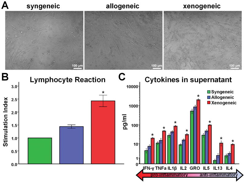
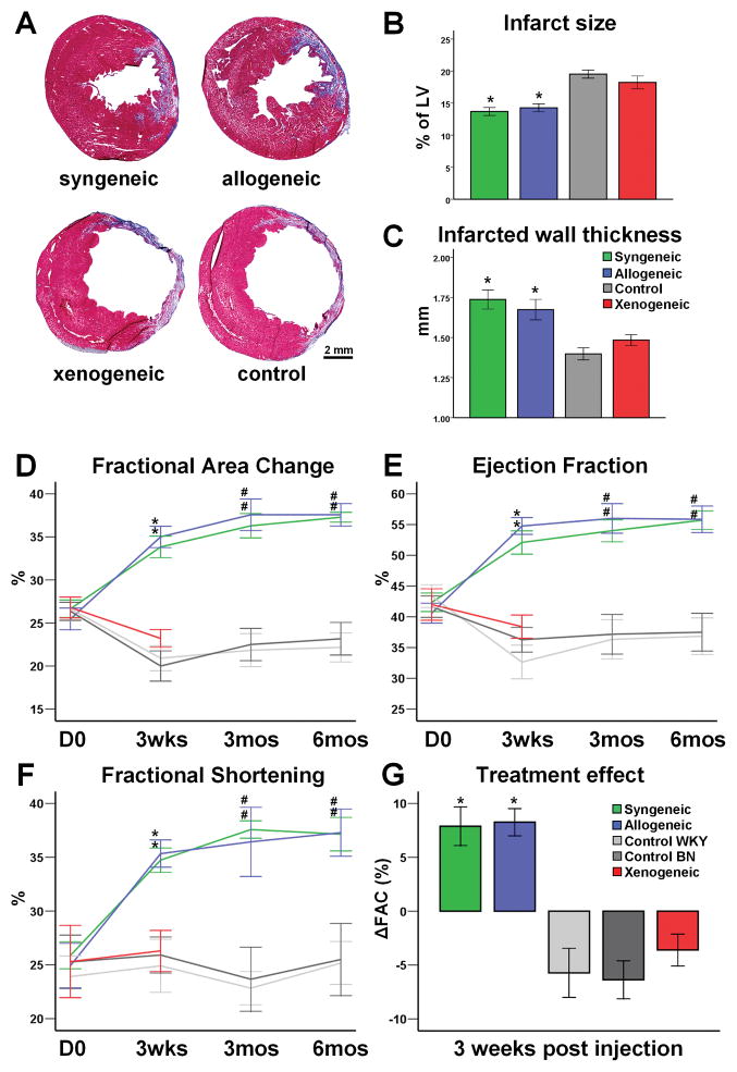
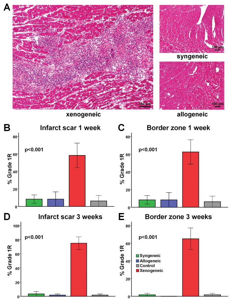
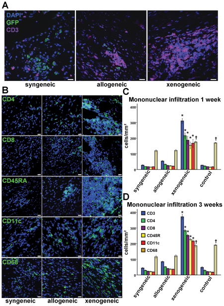
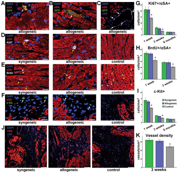
Methods and results: In vitro, CDCs expressed major histocompatibility complex class I but not class II antigens or B7 costimulatory molecules. In mixed-lymphocyte cocultures, allogeneic CDCs elicited negligible lymphocyte proliferation and inflammatory cytokine secretion. In vivo, syngeneic and allogeneic CDCs survived at similar levels in the infarcted rat heart 1 week after delivery, but few syngeneic (and even fewer allogeneic) CDCs remained at 3 weeks. Allogeneic CDCs induced a transient, mild, local immune reaction in the heart, without histologically evident rejection or systemic immunogenicity. Improvements in cardiac structure and function, sustained for 6 months, were comparable with syngeneic and allogeneic CDCs. Allogeneic CDCs stimulated endogenous regenerative mechanisms (cardiomyocyte cycling, recruitment of c-kit(+) cells, angiogenesis) and increased myocardial vascular endothelial growth factor, insulin-like growth factor-1, and hepatocyte growth factor equally with syngeneic CDCs.

Conclusions: Allogeneic CDC transplantation without immunosuppression is safe, promotes cardiac regeneration, and improves heart function in a rat myocardial infarction model, mainly through stimulation of endogenous repair mechanisms. The indirect mechanism of action rationalizes the persistence of benefit despite the evanescence of transplanted cell survival. This work motivates the testing of allogeneic human CDCs as a potential off-the-shelf product for cellular cardiomyoplasty.


Conflict of interest statement

Conflict of Interest Disclosures: EM and LM are founders and equity holders in Capricor, Inc. LM receives salary from Capricor, Inc. KM and JT receive consulting fees from Capricor, Inc. The remaining authors report no conflicts.

Figures

Figure 1
Figure 1
Phenotypic characterization of rat and human CDCs by flow cytometry. (A) Antigenic profiles of CDCs (n=4–5/group). (B) Immunophenotype of CDCs under baseline conditions and after IFN-γ stimulation (n=4–5/group). (C) CDCs at baseline express MHC class I but not MHC class II antigens. Incubation with interferon-γ upregulates expression of MHC class I and class II antigens in a time-dependent manner.
Figure 2
Figure 2
Assessment of immunogenicity of CDCs in vitro. (A) Representative images of syngeneic, allogeneic and xenogeneic cocultures. Significant lymphocyte proliferation can be observed in the xenogeneic setting. Quantitative analyses of (B) responder cell proliferation (n= 6–8/group) and (C) inflammatory cytokine secretion (n=21–26/group) demonstrate that allogeneic CDCs, contrary to xenogeneic, exhibit negligible functional immunogenicity in vitro. (* p<0.05 vs. syngeneic, allogeneic groups)
Figure 3
Figure 3
Study outline, experimental groups and CDC engraftment. (A) Study outline. (B) Experimental groups. (C) Cell engraftment by quantitative PCR 1 week (n=5–6/group) and (D) 3 weeks (n=5–6/group) post-MI and cell transplantation. Syngeneic and allogeneic CDCs demonstrated similar survival rates 1 week after transplantation, while the vast majority of xenogeneic cells had already been rejected. Three weeks post-transplantation, cell survival was poor in both syngeneic and allogeneic groups, but significantly higher after transplantation of syngeneic cells. No xenogeneic cells were detectable at 3 weeks. (* p<0.05 vs. xenogeneic group)
Figure 4
Figure 4
Structural and functional benefits following syngeneic and allogeneic CDC transplantation. (A) Representative images of Masson’s Trichrome staining of infarcted rat hearts 3 weeks post-MI. Both syngeneic and allogeneic transplantation reduced infarct size (B) and increased infarcted wall thickness (C), compared to xenogeneic or control groups (n=5–8/group). Echocardiographic assessment of LV function revealed that both syngeneic and allogeneic CDC transplantation resulted in a robust and sustained improvement of fractional area change (D), ejection fraction (E) and fractional shortening (F). The treatment effect was similar in syngeneic and allogeneic groups (G) and was sustained at least for 6 months. (* p<0.05 vs xenogeneic, control groups; # p<0.05 vs control groups; sample sizes for D–G listed in Supplemental Table 1)
Figure 5
Figure 5
Assessment of local immune rejection by H&E staining. (A) Representative images of H&E stained heart sections. No immune reaction can be detected in the allogeneic setting, while perivascular and interstitial mononuclear infiltration with no foci of myocyte damage can be observed in the xenogeneic setting (Grade 1R rejection). (B–E) Quantitative analysis of immune rejection based on the ISHLT grading system demonstrated that no significant immune rejection could be detected in the infarct scar and border zone 1 or 3 weeks after allogeneic cell transplantation. In contrast, xenogeneic cell transplantation resulted in Grade 1R rejection (n=4–5/group at each timepoint).
Figure 6
Figure 6
Assessment of local immune rejection by immunohistochemistry. (A,B) Immunohistochemistry revealed small, sparse interstitial infiltrates in the proximity of some allogeneic CDCs 3 weeks post transplantation, while large infiltrates could be detected in the xenogeneic setting. Infiltrates comprised mainly CD3+ T lymphocytes (with equal contributions of CD8+ T cytotoxic and CD4+ T helper subpopulations) and to a lesser extent CD45RA+ B lymphocytes and CD11c+ dendritic cells. CD68+ macrophages did not localize within the infiltrates and were evenly dispersed along the infarct (scale bars: 20μm). Mononuclear infiltration was significantly higher in the xenogeneic group at 1 week (C) (n=4/group) and 3 weeks (D) (n=4/group) post transplantation. (* p<0.05 vs syngeneic, control groups; † p<0.05 vs syngeneic, allogeneic groups)
Figure 7
Figure 7
Direct and indirect contributions of allogeneic CDCs to myocardial repair. Rare events of long-term engraftment and cardiogenic (A,B) or angiogenic (C) differentiation of allogeneic CDCs could be detected. More importantly, syngeneic and allogeneic CDCs promoted endogenous mechanisms of regeneration by stimulating cardiomyocyte cycling (D,E,G,H) (n=5–8/group at each timepoint), host stem cell recruitment (F,I) (n=5–8/group at each timepoint) and angiogenesis (J,K) (n=8/group). (* p<0.05 vs syngeneic, allogeneic groups; arrows in D,E denote cardiomyocyte nuclei, while arrowheads denote non-cardiomyocyte nuclei; arrows in F denote c-Kit+ cells; A–F scale bars: 20 μm; J,K scale bars: 100 μm)
Figure 8
Figure 8
Detection of beneficial paracrine factors by Western blotting. (A) Representative blots demonstrating increased secretion of VEGF, IGF1 and HGF during the first week post syngeneic and allogeneic CDC transplantation. Quantitative analysis of myocardial levels of VEGF (B), IGF-1 (C) and HGF (D) post MI (n=4–6/group at each timepoint). (* p<0.05 vs syngeneic, allogeneic groups; s: syngeneic; a: allogeneic; c: control)

References

    1. Wollert KC, Drexler H. Cell therapy for the treatment of coronary heart disease: a critical appraisal. Nat Rev Cardiol. 2010;7:204–215. - PubMed
    1. Malliaras K, Marbán E. Cardiac cell therapy: where we’ve been, where we are, and where we should be headed. Br Med Bull. 2011;98:161–185. - PMC - PubMed
    1. CADUCEUS. CArdiosphere-Derived aUtologous stem CElls to reverse ventricUlar dySfunction (NCT00893360) Available at: www.clinicaltrials.gov. - PMC - PubMed
    1. Dimmeler S, Leri A. Aging and Disease as Modifiers of Efficacy of Cell Therapy. Circ Res. 2008;102:1319–1330. - PMC - PubMed
    1. Chimenti I, Smith RR, Li TS, Gerstenblith G, Messina E, Giacomello A, Marbán E. Relative roles of direct regeneration versus paracrine effects of human cardiosphere-derived cells transplanted into infarcted mice. Circ Res. 2010;106:971–980. - PMC - PubMed

Publication types

MeSH terms