. 2012 Jan;153(1):200-11.
doi: 10.1210/en.2011-1434. Epub 2011 Nov 15.

Acute glucocorticoid administration rapidly suppresses basal and stress-induced hypothalamo-pituitary-adrenal axis activity

Affiliations

Acute glucocorticoid administration rapidly suppresses basal and stress-induced hypothalamo-pituitary-adrenal axis activity

Marcus H Andrews et al. Endocrinology. 2012 Jan.

Abstract

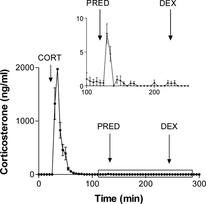
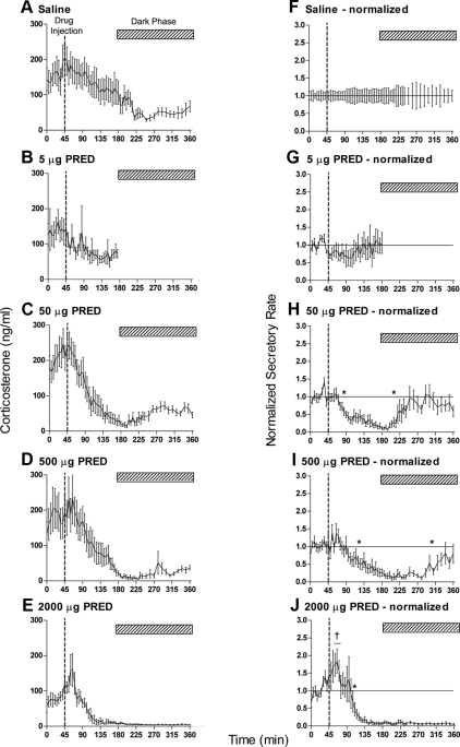
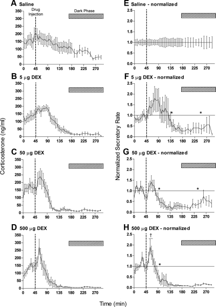
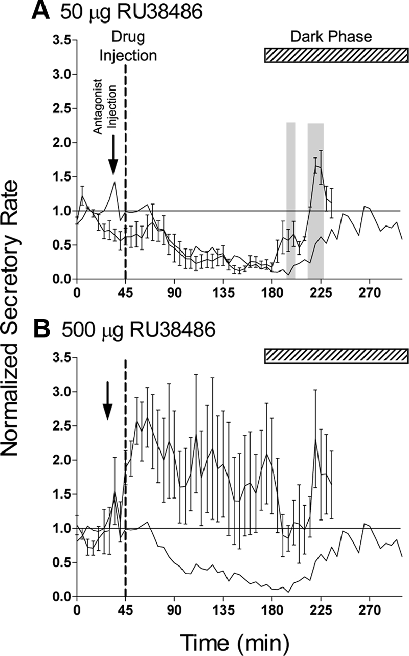
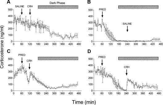
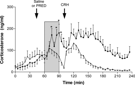
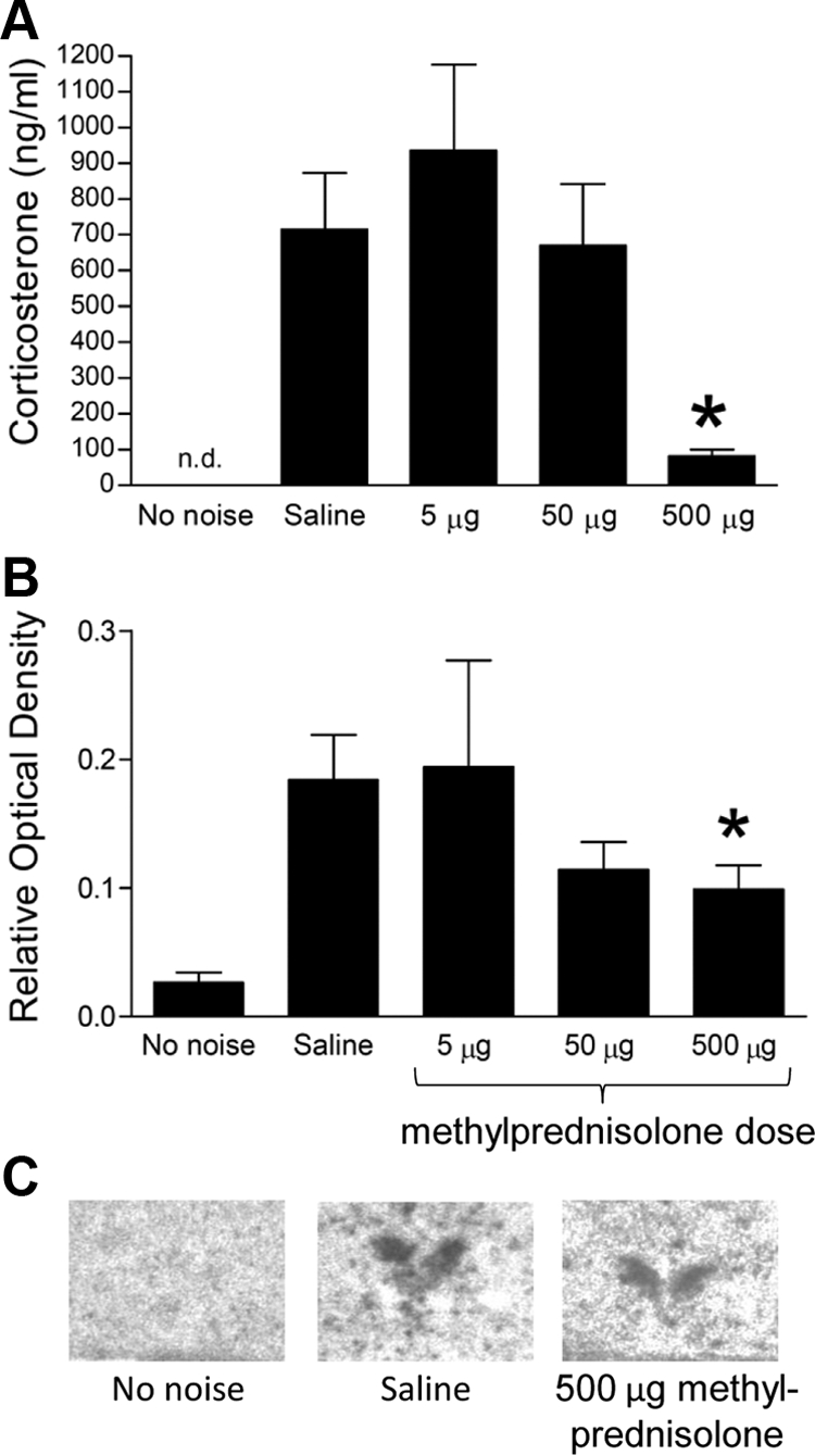
Hypothalamo-pituitary-adrenal (HPA) axis activity is subject to negative feedback control by glucocorticoids. Although the rapid component of this feedback is widely considered to contribute to regulation of dynamic HPA activity, few in vivo data exist on the temporal and pharmacological characteristics of this phenomenon. Thus, frequent automated blood sampling was undertaken in rats to determine the effects of acute glucocorticoid administration on basal and stress-induced corticosterone secretion. The glucocorticoid agonist methylprednisolone (5-2000 μg) or dexamethasone (5-500 μg) injected iv at the peak of the diurnal rhythm caused dose-dependent suppression of basal corticosterone secretion, which was attenuated by the glucocorticoid receptor antagonist RU38486. With 50 μg methylprednisolone, the onset of this suppression occurred at 40 min and remained significant for 120 min. However, although higher doses led to a greater and more sustained suppression of endogenous corticosterone, the response was delayed by the emergence of an initial stimulatory response that imposed a finite minimum delay. A corticosterone response to injection of CRH (1 μg, iv) during the period of maximal suppression indicated a suprapituitary site for the inhibitory effect glucocorticoid activation. This mechanism was supported by glucocorticoid injection immediately before a psychological stress (30 min, white noise); methylprednisolone caused dose-dependent attenuation of stress-induced corticosterone release and expression of the activity marker c-fos mRNA in the paraventricular nucleus but did not block the pituitary response to CRH. Thus, in rats, glucocorticoid receptor activation rapidly suppresses basal and stress-induced HPA activity that operates, at least in part, through a central mechanism of action.


Figures

Fig. 1.
Fig. 1.
Corticosterone clearance rates and cross-reaction of glucocorticoid agonists. Adrenalectomized animals each received iv injections of corticosterone (CORT, 400 μg), methylprednisolone (PRED, 2000 μg), and dexamethasone (DEX, 500 μg). Blood samples were assayed for corticosterone-like immunoreactivity. Inset shows the period from 100–250 min on an enlarged scale. Values represent mean ± sem (n = 7).
Fig. 2.
Fig. 2.
Acute iv injection of methylprednisolone (PRED) causes dose-dependent suppression of basal corticosterone levels. Data show the mean ± sem of the absolute hormone levels (left panels) and the data after normalization to the saline controls (right panels): A and F, Saline controls, n = 11; B and G, 5 μg, n = 8; C and H, 50 μg, n = 12; D and I, 500 μg, n = 12; E and J, 2000 μg, n = 10 (see text for normalization procedure). All injections (indicated by the broken line in each panel) were given at 1645 h when diurnal levels of circulating plasma corticosterone are high. The hatched bar in each panel indicates the dark period commencing at 1900 h. The asterisks in H–J show the first and last time points that are significantly different from saline-injected animals (P < 0.05). Note that the onset of feedback inhibition does not occur until after 40 min after methylprednisolone injection. The dagger in J indicates three successive samples that are significantly greater than the saline controls (P < 0.05). The horizontal lines in the right panels show the line of equality with saline values (i.e. normalized values equal 1).
Fig. 3.
Fig. 3.
Effects of acute iv injection of dexamethasone (DEX) on basal corticosterone levels. Data show the mean ± sem of the absolute hormone levels (left panels) and the data after normalization to the saline controls (right panels): A and E, saline controls, n = 11; B and F, 5 μg, n = 8; C and G, 50 μg, n = 6; D and H, 500 μg, n = 7. All injections (indicated by the broken line in each panel) were given at 1645 h when diurnal levels of circulating plasma corticosterone are high. The hatched bar in each panel indicates the dark period. The asterisks in F–H show the first and last time points that are significantly different from saline-injected animals (P < 0.05). Note that the onset of feedback inhibition occurs only after 50 min after DEX injection. The dagger in H indicates three successive samples that are significantly greater than the saline controls (P < 0.05). The horizontal lines in the right panels show the line of equality with saline values (i.e. normalized values equal 1). Saline control data are the same as in Fig. 2, A and E.
Fig. 4.
Fig. 4.
Effect of the GR antagonist RU38486 on the rapid inhibitory effect of iv injection of 50 μg methylprednisolone. Injection of methylprednisolone (indicated by the broken line) was preceded by injection of either 50 μg (A, n = 6) or 500 μg (B, n = 6) RU38486 (indicated by arrow). Data are the values after the normalization procedure and are mean ± sem. The solid line in each panel shows the mean response to 50 μg methylprednisolone in the absence of antagonist (data from Fig. 2H). The shaded areas in A indicate points at which the antagonist-treated data differ significantly from methylprednisolone alone (P < 0.05, repeated-measures ANOVA). The horizontal lines show the line of equality with saline values (i.e. normalized values equal 1), and the hatched bar in each panel indicates the dark period.
Fig. 5.
Fig. 5.
Effect of CRH on corticosterone secretion during methylprednisolone-induced negative feedback. A, Injection of CRH (1 μg, iv) 60 min after an injection of saline has little effect on basal HPA activity during the late light phase of the diurnal cycle (n = 4). B, Injection of methylprednisolone (PRED, 500 μg, iv) caused rapid and sustained suppression of corticosterone levels that was unaffected by subsequent injection of saline (n = 3). CRH injection was able to cause a rapid increase in corticosterone levels when given either 60 min after methylprednisolone during the maximal rate of suppression (C, n = 12) or at 240 min when corticosterone levels had reached a nadir (D, n = 4). Values are mean ± sem. Hatched bar shows the dark period. Note that for injections performed during the dark period (B and D), animals were exposed to a brief period of light.
Fig. 6.
Fig. 6.
Effect of acute glucocorticoid injection on noise stress-induced corticosterone secretion during the diurnal nadir. Single injection of methylprednisolone (PRED, 500 μg, n = 12, ▴) given 15 min before the onset of a 30-min white noise stress (104 dB, shaded area) caused a significant attenuation of the corticosterone response compared with saline-injected controls (n = 10, ■). Asterisks indicate time points that significantly differ between groups before injection of CRH (P < 0.05, repeated-measures ANOVA). Injection of CRH (1 μg, iv) at 100 min confirmed pituitary-adrenal responsiveness. Data are mean ± sem.
Fig. 7.
Fig. 7.
Glucocorticoid-induced suppression of HPA and c-fos mRNA response to noise stress. A, Injection of methylprednisolone (5–500 μg) 15 min before a 30-min noise stress significantly attenuated corticosterone levels measured from trunk blood samples obtained immediately after the end of the noise. *, P < 0.001, 500-μg dose vs. either saline or 5-μg dose (one-way ANOVA followed by post hoc Tukey's test). Corticosterone levels in animals receiving no noise were not determined (n.d.). B, Expression of c-fos mRNA in the hypothalamic PVN of animals receiving either saline or an acute dose of methylprednisolone (5–500 mg) 15 min before a 30-min noise stress. A second control group (no noise) is also included. *, P < 0.05, saline vs. 5 mg (one-way ANOVA followed by post hoc Tukey's test). C, Example autoradiograms of the PVN region in three treatment groups.

References

    1. Jingami H, Matsukura S, Numa S, Imura H. 1985. Effects of adrenalectomy and dexamethasone administration on the level of prepro-corticotropin-releasing factor messenger ribonucleic acid (mRNA) in the hypothalamus and adrenocorticotropin/β-lipotropin precursor mRNA in the pituitary in rats. Endocrinology 117:1314–1320 - PubMed
    1. Kovács KJ, Mezey E. 1987. Dexamethasone inhibits corticotropin-releasing factor gene expression in the rat paraventricular nucleus. Neuroendocrinology 46:365–368 - PubMed
    1. Drouin J, Sun YL, Nemer M. 1989. Glucocorticoid repression of pro-opiomelanocortin gene transcription. J Steroid Biochem 34:63–69 - PubMed
    1. Lightman SL, Young WS., 3rd 1989. Influence of steroids on the hypothalamic corticotropin-releasing factor and preproenkephalin mRNA responses to stress. Proc Natl Acad Sci USA 86:4306–4310 - PMC - PubMed
    1. Nakai Y, Usui T, Tsukada T, Takahashi H, Fukata J, Fukushima M, Senoo K, Imura H. 1991. Molecular mechanisms of glucocorticoid inhibition of human proopiomelanocortin gene transcription. J Steroid Biochem Mol Biol 40:301–306 - PubMed

Publication types

MeSH terms