

Biochemical and computational analysis of LNX1 interacting proteins
- PMID: 22087225
- PMCID: PMC3210812
- DOI: 10.1371/journal.pone.0026248
Biochemical and computational analysis of LNX1 interacting proteins
Abstract
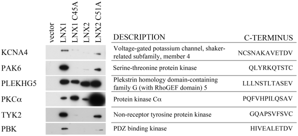
PDZ (Post-synaptic density, 95 kDa, Discs large, Zona Occludens-1) domains are protein interaction domains that bind to the carboxy-terminal amino acids of binding partners, heterodimerize with other PDZ domains, and also bind phosphoinositides. PDZ domain containing proteins are frequently involved in the assembly of multi-protein complexes and clustering of transmembrane proteins. LNX1 (Ligand of Numb, protein X 1) is a RING (Really Interesting New Gene) domain-containing E3 ubiquitin ligase that also includes four PDZ domains suggesting it functions as a scaffold for a multi-protein complex. Here we use a human protein array to identify direct LNX1 PDZ domain binding partners. Screening of 8,000 human proteins with isolated PDZ domains identified 53 potential LNX1 binding partners. We combined this set with LNX1 interacting proteins identified by other methods to assemble a list of 220 LNX1 interacting proteins. Bioinformatic analysis of this protein list was used to select interactions of interest for future studies. Using this approach we identify and confirm six novel LNX1 binding partners: KCNA4, PAK6, PLEKHG5, PKC-alpha1, TYK2 and PBK, and suggest that LNX1 functions as a signalling scaffold.
Conflict of interest statement
Figures


Similar articles
-
The Molecular and Pathophysiological Functions of Members of the LNX/PDZRN E3 Ubiquitin Ligase Family.Molecules. 2020 Dec 15;25(24):5938. doi: 10.3390/molecules25245938. Molecules. 2020. PMID: 33333989 Free PMC article. Review.
-
LNX1/LNX2 proteins: functions in neuronal signalling and beyond.Neuronal Signal. 2018 Jun 7;2(2):NS20170191. doi: 10.1042/NS20170191. eCollection 2018 Jun. Neuronal Signal. 2018. PMID: 32714586 Free PMC article. Review.
-
Proteomic analysis reveals novel ligands and substrates for LNX1 E3 ubiquitin ligase.PLoS One. 2017 Nov 9;12(11):e0187352. doi: 10.1371/journal.pone.0187352. eCollection 2017. PLoS One. 2017. PMID: 29121065 Free PMC article.
-
c-Src is a PDZ interaction partner and substrate of the E3 ubiquitin ligase Ligand-of-Numb protein X1.FEBS Lett. 2007 Oct 30;581(26):5131-6. doi: 10.1016/j.febslet.2007.09.062. Epub 2007 Oct 8. FEBS Lett. 2007. PMID: 17936276
-
Molecular evolution of the LNX gene family.BMC Evol Biol. 2011 Aug 9;11:235. doi: 10.1186/1471-2148-11-235. BMC Evol Biol. 2011. PMID: 21827680 Free PMC article.
Cited by
-
The Molecular and Pathophysiological Functions of Members of the LNX/PDZRN E3 Ubiquitin Ligase Family.Molecules. 2020 Dec 15;25(24):5938. doi: 10.3390/molecules25245938. Molecules. 2020. PMID: 33333989 Free PMC article. Review.
-
LNX1 Contributes to Cell Cycle Progression and Cisplatin Resistance.Cancers (Basel). 2021 Aug 12;13(16):4066. doi: 10.3390/cancers13164066. Cancers (Basel). 2021. PMID: 34439220 Free PMC article.
-
LNX1/LNX2 proteins: functions in neuronal signalling and beyond.Neuronal Signal. 2018 Jun 7;2(2):NS20170191. doi: 10.1042/NS20170191. eCollection 2018 Jun. Neuronal Signal. 2018. PMID: 32714586 Free PMC article. Review.
-
Scaffold Protein Lnx1 Stabilizes EphB Receptor Kinases for Synaptogenesis.Front Mol Neurosci. 2022 Apr 21;15:861873. doi: 10.3389/fnmol.2022.861873. eCollection 2022. Front Mol Neurosci. 2022. PMID: 35531068 Free PMC article.
-
Is IIIG9 a New Protein with Exclusive Ciliary Function? Analysis of Its Potential Role in Cancer and Other Pathologies.Cells. 2022 Oct 21;11(20):3327. doi: 10.3390/cells11203327. Cells. 2022. PMID: 36291193 Free PMC article. Review.
References
-
- Doyle DA, Lee A, Lewis J, Kim E, Sheng M, et al. Crystal structures of a complexed and peptide-free membrane protein-binding domain: molecular basis of peptide recognition by PDZ. Cell. 1996;85(7):1067–1076. - PubMed
-
- Tochio H, Mok YK, Zhang Q, Kan HM, Bredt DS, et al. Formation of nNOS/PSD-95 PDZ dimer requires a preformed beta-finger structure from the nNOS PDZ domain. J Mol Biol. 2000;303(3):359–370. - PubMed
-
- Wang P, Zhang Q, Tochio H, Fan JS, Zhang M. Formation of a native-like beta-hairpin finger structure of a peptide from the extended PDZ domain of neuronal nitric oxide synthase in aqueous solution. Eur J Biochem. 2000;267(11):3116–3122. - PubMed
-
- Zimmermann P. The prevalence and significance of PDZ domain-phosphoinositide interactions. Biochim Biophys Acta. 2006;1761(8):947–956. - PubMed
-
- Wang T, Montell C. Phototransduction and retinal degeneration in Drosophila. Pflugers Arch. 2007;454(5):821–847. - PubMed
Publication types
MeSH terms
Substances
Grants and funding
LinkOut - more resources
Full Text Sources
