Comparative Study
. 2011;6(11):e26580.
doi: 10.1371/journal.pone.0026580. Epub 2011 Nov 8.

Candida albicans yeast and hyphae are discriminated by MAPK signaling in vaginal epithelial cells

Affiliations
Comparative Study

Candida albicans yeast and hyphae are discriminated by MAPK signaling in vaginal epithelial cells

David L Moyes et al. PLoS One. 2011.

Abstract

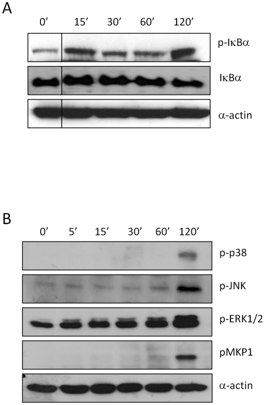
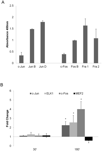
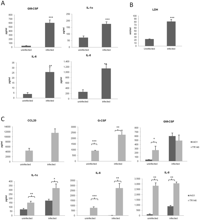
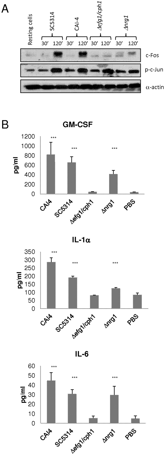
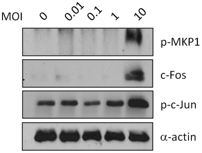
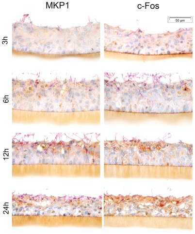
We previously reported that a bi-phasic innate immune MAPK response, constituting activation of the mitogen-activated protein kinase (MAPK) phosphatase MKP1 and c-Fos transcription factor, discriminates between the yeast and hyphal forms of Candida albicans in oral epithelial cells (ECs). Since the vast majority of mucosal Candida infections are vaginal, we sought to determine whether a similar bi-phasic MAPK-based immune response was activated by C. albicans in vaginal ECs. Here, we demonstrate that vaginal ECs orchestrate an innate response to C. albicans via NF-κB and MAPK signaling pathways. However, unlike in oral ECs, the first MAPK response, defined by c-Jun transcription factor activation, is delayed until 2 h in vaginal ECs but is still independent of hypha formation. The 'second' or 'late' MAPK response, constituting MKP1 and c-Fos transcription factor activation, is identical to oral ECs and is dependent upon both hypha formation and fungal burdens. NF-κB activation is immediate but independent of morphology. Furthermore, the proinflammatory response in vaginal ECs is different to oral ECs, with an absence of G-CSF and CCL20 and low level IL-6 production. Therefore, differences exist in how C. albicans activates signaling mechanisms in oral and vaginal ECs; however, the activation of MAPK-based pathways that discriminate between yeast and hyphal forms is retained between these mucosal sites. We conclude that this MAPK-based signaling pathway is a common mechanism enabling different human epithelial tissues to orchestrate innate immune responses specifically against C. albicans hyphae.


Conflict of interest statement

Competing Interests: The authors have declared that no competing interests exist.

Figures

Figure 1
Figure 1. C. albicans infection of A431 epithelial cells activates NF-κB and MAPK signaling.
C. albicans SC5314 was added to A431 vaginal ECs under standard culture conditions for 5, 15, 30, 60 min and 2 h. Total protein was isolated and phosphorylation of (A) IκBα or (B) p38, JNK, ERK1/2 and MKP1 were assessed by Western blotting. Bands are shown with an α-actin loading control. A C. albicans:EC MOI of 10∶1 was used. Data are representative of three independent experiments.
Figure 2
Figure 2. Activation of MAPK transcription factors by C. albicans in A431 vaginal epithelial cells.
(A) Resting levels of DNA binding activity (absorbance values) of AP-1 transcription factor members in nuclear extracts from A431 ECs, measured by TransAm ELISA. (B) Changes in DNA binding activity of c-Jun, Elk1, c-Fos, and MEF2 in nuclear extracts of A431 ECs 30 and 180 min post-infection with C. albicans SC5314 by TransAm ELISA. These MAPK-activated transcription factors have previously been identified as showing altered DNA binding activity in oral ECs. Data are represented as fold change relative to resting levels at 0 h. A C. albicans:EC MOI of 10∶1 was used. Data represent mean values ± SEM and are representative of a minimum of three independent experiments. Statistical analysis (B) of raw data for infected versus uninfected cells was performed using the unpaired, two-tailed t-test * p<0.05.
Figure 3
Figure 3. Cytokine activation and cell damage induced by C. albicans in A431 vaginal epithelial cells and TR146 oral epithelial cells.
(A) C. albicans SC5314 was added to monolayers of A431 ECs for 24 h and the cell culture medium collected and assessed for cytokine proteins by multiplex microbead assay (luminex) or ELISA (CCL20). (B) LDH (lactate dehydrogenase) release (measure of cell damage) from A431 ECs 24 h post-infection with C. albicans SC5314. (C) Comparison of cytokine protein release by A431 ECs and TR146 ECs 24 h post-infection with C. albicans SC5314. A C. albicans:EC MOI of 0.01 was used for all the experiments. Data are mean values ± SEM of three independent experiments. Statistical analysis of infected versus uninfected epithelial cells (A & B) or oral versus vaginal epithelial cells (C) was performed using the unpaired, two-tailed t-test. * p<0.05, ** p<0.01, *** p<0.001.
Figure 4
Figure 4. Induction of c-Jun phosphorylation, c-Fos and cytokine production in A431 vaginal epithelial cells is dependent on C. albicans hypha formation.
(A) C. albicans SC5314 (wild type), CAI4 (parent control), Δefg1/cph1 (non-filamentous) and Δnrg1 (hyperfilamentous) were added to A431 vaginal ECs under standard culture conditions for 30 min and 2 h. Total protein was isolated and induction of c-Jun phosphorylation and c-Fos assessed by Western blotting. Bands are shown alongside an α-actin loading control. (B) Cytokine protein production by A431 ECs 24 h post-infection with all four strains as measured by multiplex microbead assay (luminex). A C. albicans:EC MOI of 10∶1 (A) and 0.01 (B) was used. Data are representative (A) or mean ± SEM (B) of three independent experiments. Statistical analysis (B) of SC5314, CAI-4, Δefg1/cph1 and Δnrg1 infected versus PBS-treated controls was performed using the ANOVA test with Bonferroni post-hoc analysis. *** = p<0.001.
Figure 5
Figure 5. Effect of fungal burdens on MKP1, c-Fos and c-Jun activation.
A431 vaginal ECs were infected for 2 h with C. albicans SC5314 at MOI's ranging between 0.01 and 10. Total protein was isolated and phosphorylation of MKP1 and c-Jun and c-Fos induction was assessed by Western blotting. Bands are shown alongside an α-actin loading control. Data are representative of four independent experiments.
Figure 6
Figure 6. Expression of MKP1 and c-Fos in vaginal RHE.
Increase in MKP1 and c-Fos expression in vaginal epithelium is associated with C. albicans SC5314 hypha formation, with minimal activation at the surface by 3 h and gradually increased activation co-localising in the epithelial layers where hyphae penetrate and invade at 6, 12 and 24 h (dark brown staining). Resting levels of MKP1 and c-Fos expression can be seen in areas without C. albicans at each time point (sectional control).

References

    1. Sobel JD. Pathogenesis and epidemiology of vulvovaginal candidiasis. Annals N Y Acad Sci. 1988;544:547–557. - PubMed
    1. Moyes DL, Runglall M, Murciano C, Shen C, Nayar D, et al. A biphasic innate immune MAPK response discriminates between the yeast and hyphal forms of Candida albicans in epithelial cells. Cell Host Microbe. 2010;8:225–235. - PMC - PubMed
    1. Weindl G, Naglik JR, Kaesler S, Biedermann T, Hube B, et al. Human epithelial cells establish direct antifungal defense through TLR4-mediated signaling. J Clin Invest. 2007;117:3664–3672. - PMC - PubMed
    1. Fidel PL, Jr, Barousse M, Espinosa T, Ficarra M, Sturtevant J, et al. An intravaginal live Candida challenge in humans leads to new hypotheses for the immunopathogenesis of vulvovaginal candidiasis. Infect Immun. 2004;72:2939–2946. - PMC - PubMed
    1. Naglik JR, Moyes D, Makwana J, Kanzaria P, Tsichlaki E, et al. Quantitative expression of the Candida albicans secreted aspartyl proteinase gene family in human oral and vaginal candidiasis. Microbiology. 2008;154:3266–3280. - PMC - PubMed

Publication types