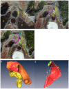
Visualization of the left extraperitoneal space and spatial relationships to its related spaces by the visible human project
- PMID: 22087259
- PMCID: PMC3210141
- DOI: 10.1371/journal.pone.0027166
Visualization of the left extraperitoneal space and spatial relationships to its related spaces by the visible human project
Abstract
Background: The major hindrance to multidetector CT imaging of the left extraperitoneal space (LES), and the detailed spatial relationships to its related spaces, is that there is no obvious density difference between them. Traditional gross anatomy and thick-slice sectional anatomy imagery are also insufficient to show the anatomic features of this narrow space in three-dimensions (3D). To overcome these obstacles, we used a new method to visualize the anatomic features of the LES and its spatial associations with related spaces, in random sections and in 3D.
Methods: In conjunction with Mimics® and Amira® software, we used thin-slice cross-sectional images of the upper abdomen, retrieved from the Chinese and American Visible Human dataset and the Chinese Virtual Human dataset, to display anatomic features of the LES and spatial relationships of the LES to its related spaces, especially the gastric bare area. The anatomic location of the LES was presented on 3D sections reconstructed from CVH2 images and CT images.
Principal findings: What calls for special attention of our results is the LES consists of the left sub-diaphragmatic fat space and gastric bare area. The appearance of the fat pad at the cardiac notch contributes to converting the shape of the anteroexternal surface of the LES from triangular to trapezoidal. Moreover, the LES is adjacent to the lesser omentum and the hepatic bare area in the anterointernal and right rear direction, respectively.
Conclusion: The LES and its related spaces were imaged in 3D using visualization technique for the first time. This technique is a promising new method for exploring detailed communication relationships among other abdominal spaces, and will promote research on the dynamic extension of abdominal diseases, such as acute pancreatitis and intra-abdominal carcinomatosis.
Conflict of interest statement
Figures


Similar articles
-
Anatomic pathways of peripancreatic fluid draining to mediastinum in recurrent acute pancreatitis: visible human project and CT study.PLoS One. 2013 Apr 17;8(4):e62025. doi: 10.1371/journal.pone.0062025. Print 2013. PLoS One. 2013. PMID: 23614005 Free PMC article.
-
Visualization of the omental bursa and its spatial relationships to left subphrenic extraperitoneal spaces by the second Chinese Visible Human model.Surg Radiol Anat. 2015 Jul;37(5):473-81. doi: 10.1007/s00276-015-1462-3. Epub 2015 Mar 28. Surg Radiol Anat. 2015. PMID: 25820977 Free PMC article.
-
[Cross-section anatomy of the subphrenic spaces].Zhonghua Wai Ke Za Zhi. 1996 Feb;34(2):120-2. Zhonghua Wai Ke Za Zhi. 1996. PMID: 9388340 Chinese.
-
Anatomic CT demonstration of the peritoneal spaces, ligaments, and mesenteries: normal and pathologic processes.Radiographics. 1995 Jul;15(4):755-70. doi: 10.1148/radiographics.15.4.7569127. Radiographics. 1995. PMID: 7569127 Review.
-
[Echographic anatomy of the greater peritoneal cavity and its recesses].Radiol Med. 1988 Jan-Feb;75(1-2):46-55. Radiol Med. 1988. PMID: 3279472 Review. Italian.
Cited by
-
Anatomic pathways of peripancreatic fluid draining to mediastinum in recurrent acute pancreatitis: visible human project and CT study.PLoS One. 2013 Apr 17;8(4):e62025. doi: 10.1371/journal.pone.0062025. Print 2013. PLoS One. 2013. PMID: 23614005 Free PMC article.
-
Visualization of Penile Suspensory Ligamentous System Based on Visible Human Data Sets.Med Sci Monit. 2017 May 22;23:2436-2444. doi: 10.12659/msm.901926. Med Sci Monit. 2017. PMID: 28530218 Free PMC article.
-
Retrocrural space involvement on computed tomography as a predictor of mortality and disease severity in acute pancreatitis.PLoS One. 2014 Sep 15;9(9):e107378. doi: 10.1371/journal.pone.0107378. eCollection 2014. PLoS One. 2014. PMID: 25222846 Free PMC article.
-
Visualization of the omental bursa and its spatial relationships to left subphrenic extraperitoneal spaces by the second Chinese Visible Human model.Surg Radiol Anat. 2015 Jul;37(5):473-81. doi: 10.1007/s00276-015-1462-3. Epub 2015 Mar 28. Surg Radiol Anat. 2015. PMID: 25820977 Free PMC article.
-
Pancreaticopleural fistula in children: Report of 2 cases.Radiol Case Rep. 2022 Jan 18;17(3):987-990. doi: 10.1016/j.radcr.2022.01.007. eCollection 2022 Mar. Radiol Case Rep. 2022. PMID: 35106110 Free PMC article.
References
-
- Rowe PM. Visible Human Project pays back investment. Lancet. 1999;353:46. - PubMed
-
- Xia CL, Xu L, Xu J, Ju WN, Piao HH, et al. Three-dimensional reconstruction of internal capsule and its clinical application. Journal of Jilin University (Medicine Edition) 2007;33:898–900.
-
- Chen R, Zhang S, Zhang W, Tan L, Li Q, et al. A comparative study of thin-layer cross-sectional anatomic morphology and CT images of the basal cistern and its application in acute craniocerebral traumas. Surg Radiol Anat. 2009;31:129–138. - PubMed
-
- Wu Y, Zhang SX, Luo N, Qiu MG, Tan LW, et al. Creation of the digital three-dimensional model of the prostate and its adjacent structures based on Chinese visible human. Surg Radiol Anat. 2010;32:629–635. - PubMed
Publication types
MeSH terms
LinkOut - more resources
Full Text Sources

