. 2011;6(11):e27469.
doi: 10.1371/journal.pone.0027469. Epub 2011 Nov 8.

The role of purinergic signaling on deformation induced injury and repair responses of alveolar epithelial cells

Affiliations

The role of purinergic signaling on deformation induced injury and repair responses of alveolar epithelial cells

Hewan A Belete et al. PLoS One. 2011.

Abstract

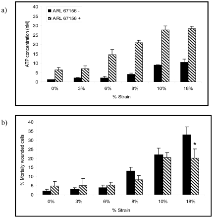
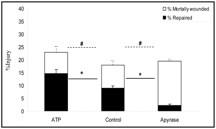
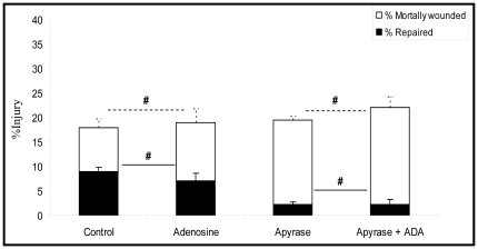
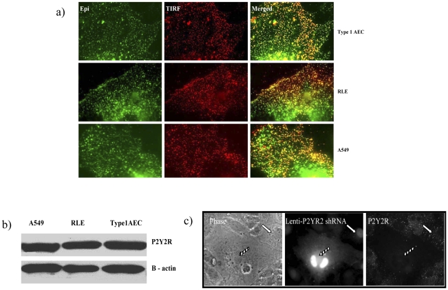
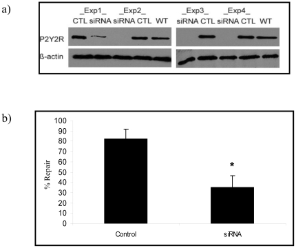
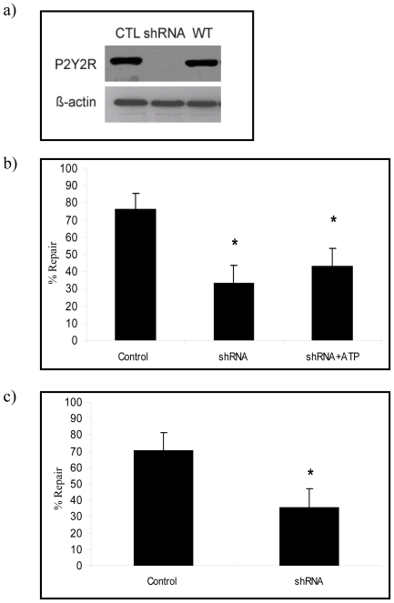
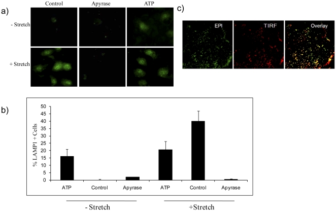
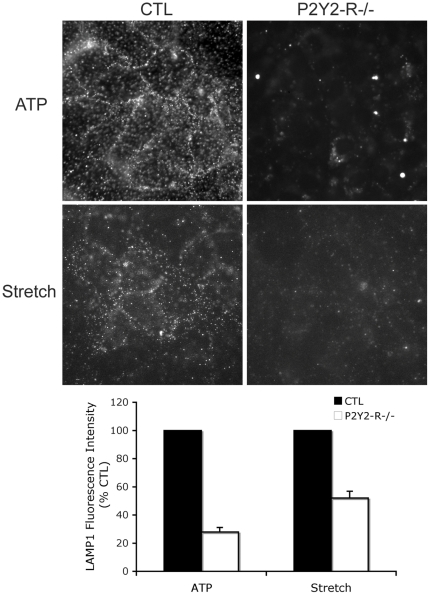
Cell wounding is an important driver of the innate immune response of ventilator-injured lungs. We had previously shown that the majority of wounded alveolus resident cells repair and survive deformation induced insults. This is important insofar as wounded and repaired cells may contribute to injurious deformation responses commonly referred to as biotrauma. The central hypothesis of this communication states that extracellular adenosine-5' triphosphate (ATP) promotes the repair of wounded alveolus resident cells by a P2Y2-Receptor dependent mechanism. Using primary type 1 alveolar epithelial rat cell models subjected to micropuncture injury and/or deforming stress we show that 1) stretch causes a dose dependent increase in cell injury and ATP media concentrations; 2) enzymatic depletion of extracellular ATP reduces the probability of stretch induced wound repair; 3) enriching extracellular ATP concentrations facilitates wound repair; 4) purinergic effects on cell repair are mediated by ATP and not by one of its metabolites; and 5) ATP mediated cell salvage depends at least in part on P2Y2-R activation. While rescuing cells from wounding induced death may seem appealing, it is possible that survivors of membrane wounding become governors of a sustained pro-inflammatory state and thereby perpetuate and worsen organ function in the early stages of lung injury syndromes. Means to uncouple P2Y2-R mediated cytoprotection from P2Y2-R mediated inflammation and to test the preclinical efficacy of such an undertaking deserve to be explored.


Conflict of interest statement

Competing Interests: The authors have declared that no competing interests exist.

Figures

Figure 1
Figure 1. Effect of strain amplitude on medium ATP concentration and % mortally wounded type 1 AECs.
Type 1 AECs grown on Flexcell culture plates were stretched in the presence (hatched bars) or absence (black bars) of the ectoenzyme inhibitor ARL 67156 (100 µM) for 10 minutes (frequency = 0.5 Hz and strain rate = 20% sec−1). ATP concentration in supernatant was measured using a luciferase assay. (a) Plot of strain amplitude, defined as the % radial length change during each stretch cycle vs. nanomolar ATP media concentration; (b) Plot of strain amplitude vs. % mortally wounded cells as indicated by PI labeling (n = 6). (*  =  p<0.05). Data are presented as means ± one standard error of the mean.
Figure 2
Figure 2. Effects of extracellular ATP and Apyrase on % injury and % repair of type 1 AECs.
Apyrase (20 U/ml) treated, ATP (10 µM) treated and control cells were subjected to cyclic stretch of 10% for 10 minutes (Frequency = 0.5 Hz strain rate = 20% sec−1) in the presence of FDx. PI labeling was used to identify mortally wounded cells. % injured, % repaired and % mortally wounded rates were calculated according to the formula described in the methods section. The y axis represents the percentage of cells that were injured as a result of stretch (% injury). The white bar represents cells that were injured & repaired, while the black bar represents mortally wounded cells, i.e. cells that were injured but failed to repair (n = 6) (*  =  p<0.05, # p>0.05). Data are presented as means ± one standard error of the mean.
Figure 3
Figure 3. Effects of adenosine and adenosine deaminase on % injury and % repair of type 1 AECs.
Adenosine treated, adenosine deaminase + apyrase treated and control cells were subjected to cyclic stretch of 10% for 10 minutes (Frequency = 0.5 Hz strain rate = 20% sec−1) in the presence of FDx. PI labeling was used to identify mortally wounded cells. % injured, % repaired and % mortally wounded rates were calculated according to the formula described in the methods section (n = 6) (# p>0.05) Data are presented as means ± one standard error of the mean.
Figure 4
Figure 4. P2Y2-R expression in type 1 AEC, A549 and RLE cells.
(a) Representative Epi and TIRF images of type 1 AECs, A549 and RLE immunostained with anti-P2Y2-R. TIRF mode (red) and EPI mode images were overlaid (yellow) to show colocalization. (b) Protein abundance of P2Y2-R (top) in relation to loading control, beta actin (bottom), in type 1 AEC, A549 and RLE cells. (c) Immunostaining showing P2Y2-R null (gridded arrows) and P2Y2-R positive (white arrow) type 1 AEC. Left panel shows phase images of type 1 AEC culture; middle panel shows GFP fluorescence as a result of lenti-P2Y2-R shRNA infection; right panel shows expression of P2Y2-R. Note the absence of P2Y2-R immunolabel in the null cell (gridded arrow) compared to the surrounding uninfected wild type control cells.
Figure 5
Figure 5. Effect of P2Y2-R silencing (via siRNA) on PM wound repair in A549 cells.
(a) P2Y2R protein expression in P2Y2R-targeted siRNA transfected A549 cells (siRNA) compared to those transfected with scrambled control (CTL) and wild-type A549 (WT) (top). Beta-actin was used as a loading control (bottom). Western blots shown are results of four separate experiments (Exp1-4) (b) A549 cells transfected with P2Y2R-targeted siRNA (siRNA) or non-targeting scrambled control siRNA (control) were subjected to micropuncture injury. % repair was calculated according to the formula described in the methods section (n = 17 cells/ group) (* indicates p<0.05). Data are presented as means ± one standard error of the mean.
Figure 6
Figure 6. Effect of P2Y2-R knockdown (via shRNA) on PM wound repair in RLE and Type 1 AECs.
(a) P2Y2R protein expression in P2Y2R-shRNA lentivirus infected RLE cells (shRNA) compared to those that were infected with non-targeting shRNA lentiviral particles (CTL) and wild type RLE cells (WT) (top). Beta-actin was used as a loading control (bottom). (b) P2Y2R null (shRNA) and P2Y2R positive RLE (control) were subjected to micropuncture injury in the presence or absence of ATP and % repair was calculated according to the formula described in the methods section (n = 21 cells/group). (c) P2Y2R null (gridded arrows) and P2Y2R positive (white arrow) type 1 AECs were subjected to micropuncture injury and % repair was calculated according to the formula described in the methods section (n = 17 cells/ group) (* indicates p<0.05). Data are presented as means ± one standard error of the mean.
Figure 7
Figure 7. Surface LAMP1 expression in live type 1 AECs.
(a) Epifluorescence images of LAMP1 immunostaining in type 1 AEC. Cells were treated with 10 µM of ATP or 20 U/ml Apyrase in the presence or absence of stretch (amplitude = 6% frequency = 0.5 Hz strain rate = 20%sec−1 t = 8 min) and were labeled with LAMP-1 antibody at 4°C. (b) Percentages of LAMP1 positive cells were quantified by dividing the number of LAMP1 positive cells by the total number of cells in each image field. Data are presented as means ± one standard error of the mean. (c) TIRF (red) and EPI fluorescence images were overlaid (yellow) to validate the expression of LAMP1 at the PM.
Figure 8
Figure 8. Plasma membrane LAMP1 in wild type (CTL) and P2Y2R −/− RLE cells.
Cells were either treated with 50 µM ATP (top row, n = 5) or stretched (middle row; amplitude = 10% frequency = 0.5Hz strain rate = 40%sec−1 t = 8 min, n = 4) and subsequently labeled with LAMP-1 antibody at 4°C. Fluorescence intensity of P2Y2-R −/− RLE cells was expressed as % of the corresponding wild type CTL and data presented as means ± one standard error of the mean (bottom row).

References

    1. Shen SS, Tucker WC, Chapman ER, Steinhardt RA. Molecular regulation of membrane resealing in 3T3 fibroblasts. J Biol Chem. 2005;280:1652–1660. - PubMed
    1. Dreyfuss D, Saumon G. Ventilator-induced lung injury: lessons from experimental studies. Am J Respir Crit Care Med. 1998;157:294–323. - PubMed
    1. Vlahakis NE, Hubmayr RD. Cellular stress failure in ventilator-injured lungs. Am J Respir Crit Care Med. 2005;171:1328–1342. - PMC - PubMed
    1. Gattinoni L, Carlesso E, Cressoni M. Assessing gas exchange in acute lung injury/acute respiratory distress syndrome: diagnostic techniques and prognostic relevance. Curr Opin Crit Care. 2011;17:18–23. - PubMed
    1. Wheeler AP, Bernard GR. Acute lung injury and the acute respiratory distress syndrome: a clinical review. Lancet. 2007;369:1553–1564. - PubMed

Publication types

Substances

LinkOut - more resources