Review
. 2012 Jun;22(3):372-82.
doi: 10.1016/j.conb.2011.10.019. Epub 2011 Nov 14.

Nanoscale analysis of structural synaptic plasticity

Affiliations
Review

Nanoscale analysis of structural synaptic plasticity

Jennifer N Bourne et al. Curr Opin Neurobiol. 2012 Jun.

Abstract

Structural plasticity of dendritic spines and synapses is an essential mechanism to sustain long lasting changes in the brain with learning and experience. The use of electron microscopy over the last several decades has advanced our understanding of the magnitude and extent of structural plasticity at a nanoscale resolution. In particular, serial section electron microscopy (ssEM) provides accurate measurements of plasticity-related changes in synaptic size and density and distribution of key cellular resources such as polyribosomes, smooth endoplasmic reticulum, and synaptic vesicles. Careful attention to experimental and analytical approaches ensures correct interpretation of ultrastructural data and has begun to reveal the degree to which synapses undergo structural remodeling in response to physiological plasticity.


Figures

Figure 1
Figure 1
Perisynaptic astroglial processes are intact in acute hippocampal slices maintained in an interface chamber, but are lost or retracted when slices are maintained under submersion conditions. Astroglial processes are colorized in blue. (A) EM from perfusion fixed hippocampus. (Ai) A region enlarged from A that highlights the presence of perisynaptic astroglial at a spine synapse. (B) EM from a hippocampal slice maintained in an interface chamber showing numerous astroglial processes. (Bi) Close up of (B) showing an astroglial process contacting the neck of a dendritic spine. (C) EM from a hippocampal slice recovered in a submersion chamber showing the absence of astroglial processes resulting in an expansion of extracellular space. (Ci) Close up of (C) demonstrating a dendrite and synapse with no perisynaptic astroglial processes. Scale bars = 0.5 microns.
Figure 2
Figure 2
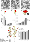
Temperature during slice preparation ultimately influences total synapse number in the recovered slices. Tissue quality of hippocampal neuropil in stratum radiatum of area CA1 is comparable across (A) slices prepared under ice-cold conditions and maintained at ~32° C for 9–10 hr in an interface chamber, (B) hippocampus perfusion-fixed in vivo, and (C) in slices prepared at room temperature and maintained at ~32° C for 4.5–5.5 hr in an interface chamber. (D) Three-dimensional reconstructions of dendrites from each of the three conditions. Scale cube = 1 micron on a side. (E) Dendrites from ice-cold slice preparations had a higher spine density at 9–10 hr in vitro, compared to perfusion-fixed hippocampus and room temperature slices (P<0.02). Adapted from Bourne et al., 2007.
Figure 3
Figure 3
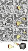
Serial images through two spines illustrate the need for ssEM to identify the location of PSDs and polyribosomes. (A–C) Serial images illustrating the presence (black arrows) of polyribosomes on some sections and their absence in the same locations on adjacent sections (white arrows). (Ai-Ci) Same images with dendritic spines highlighted in yellow, PSDs in red and polyribosomes in magenta Scale bar = 0.5 microns for both columns. (D) Three-dimensional reconstruction of spines (yellow), PSDs (red) and polyribosomes (magenta). Scale cube = 0.5 microns on a side.
Figure 4
Figure 4
Dramatic structural synaptic scaling revealed by ssEM following induction of LTP by TBS in the mature hippocampus. (A) EM and (Ai) reconstruction of a small thin spine (category T, head diameter <0.45 μm). (B) EM and (Bi) reconstruction of a medium thin spine (category T, head diameter >0.45 μm but <0.6 μm). (C) EM and (Ci) reconstruction of a mushroom spine (M, head diameter >0.6 μm). D. Small thin spines were significantly reduced in number (p<0.05) while (E) PSDs on all remaining spines were significantly enlarged (**p<0.01; *p<0.05). (F) Reconstructions of control and LTP dendrites demonstrate equal summed PSD area per micron length of dendrite, despite large differences in their average synapse size and density. Scale cube = 0.5 microns on a side. (G) Average summed PSD area per micron length of dendrite is the same for dendrites across all times in the control and TBS-LTP conditions. Adapted from Bourne and Harris, 2011.

References

    1. Palay SL, Palade GE. The fine structure of neurons. J Biophys Biochem Cytol. 1955;1:69–88. - PMC - PubMed
    1. Gray EG. Axo-somatic and axo-dendritic synapses of the cerebral cortex: an electron microscope study. J Anat. 1959;93:420–433. - PMC - PubMed
    1. Bourne JN, Sorra KE, Hurlburt J, Harris KM. Polyribosomes are increased in spines of CA1 dendrites 2 h after the induction of LTP in mature rat hippocampal slices. Hippocampus. 2007;17:1–4. - PubMed
    1. Bourne JN, Harris KM. Coordination of size and number of excitatory and inhibitory synapses results in a balanced structural plasticity along mature hippocampal CA1 dendrites during LTP. Hippocampus. 2011;21:354–373. Three-dimensional analyses of dendritic segments revealed significant structural plasticity of both excitatory and inhibitory synapses following the induction of LTP with TBS. Turnover of spines within t Three-dimensional analyses of dendritic segments revealed significant structural plasticity of both excitatory and inhibitory synapses following the induction of LTP with TBS. Turnover of spines within the first 30 minutes led to a decrease in small thin spines by 2 hours that was perfectly counterbalanced by an increase in PSD area, such that the summed PSD area per unit micron length of dendrite remained constant between control and LTP conditions. These data suggest that structural synaptic scaling may be an important cellular mechanism of plasticity in the mature hippocampus.he first 30 minutes led to a decrease in small thin spines by 2 hours that was perfectly counterbalanced by an increase in PSD area, such that the summed PSD area per unit micron length of dendrite remained constant between control and LTP conditions. These data suggest that structural synaptic scaling may be an important cellular mechanism of plasticity in the mature hippocampus. - PMC - PubMed
    1. Fiala JC, Allwardt B, Harris KM. Dendritic spines do not split during hippocampal LTP or maturation. Nat Neurosci. 2002;5:297–298. - PubMed