

Foreskin T-cell subsets differ substantially from blood with respect to HIV co-receptor expression, inflammatory profile, and memory status
- PMID: 22089029
- PMCID: PMC3288185
- DOI: 10.1038/mi.2011.56
Foreskin T-cell subsets differ substantially from blood with respect to HIV co-receptor expression, inflammatory profile, and memory status
Abstract
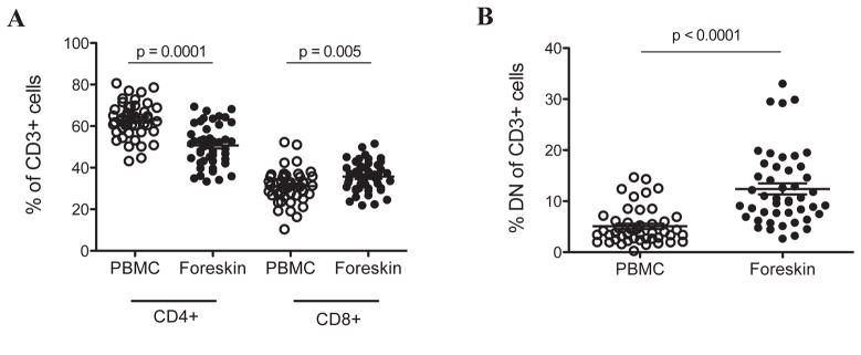
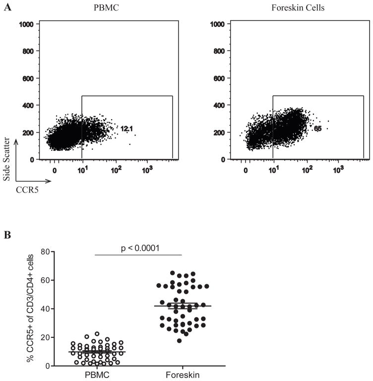
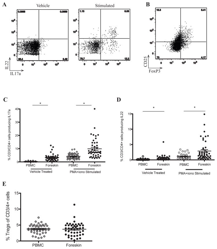
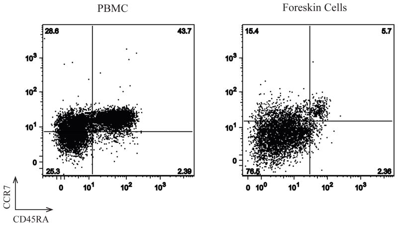
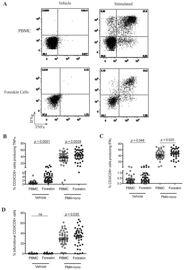
The foreskin is the main site of heterosexual human immunodeficiency virus (HIV) acquisition in uncircumcised men, but functional data regarding T-cell subsets present at this site are lacking. Foreskin tissue and blood were obtained from Ugandan men undergoing elective adult circumcision. Tissue was treated by mechanical and enzymatic digestion followed by T-cell subset identification and assessment of cytokine production using flow cytometry. Foreskin CD4(+) T cells were predominantly an effector memory phenotype, and compared with blood they displayed a higher frequency of CCR5 expression (42.0% vs. 9.9%) and interleukin-17 production. There was no difference in T-regulatory cell frequency, but interferon-γ and tumor necrosis factor-α production were increased in foreskin CD8(+) T cells. These novel techniques demonstrate that the foreskin represents a proinflammatory milieu that is enriched for HIV-susceptible T-cell subsets. Further characterization of foreskin T-cell subsets may help to define the correlates of HIV susceptibility in the foreskin.
Conflict of interest statement
The authors have nothing to disclose and no conflicts of interest.
Figures
Similar articles
-
Characterization of CD4+ T cell subsets and HIV susceptibility in the inner and outer foreskin of Ugandan men.Am J Reprod Immunol. 2019 Jul;82(1):e13143. doi: 10.1111/aji.13143. Epub 2019 May 24. Am J Reprod Immunol. 2019. PMID: 31081958
-
Immune correlates of HIV exposure without infection in foreskins of men from Rakai, Uganda.Mucosal Immunol. 2014 May;7(3):634-44. doi: 10.1038/mi.2013.83. Epub 2013 Oct 23. Mucosal Immunol. 2014. PMID: 24150258 Free PMC article.
-
Schistosoma mansoni Infection in Ugandan Men Is Associated with Increased Abundance and Function of HIV Target Cells in Blood, but Not the Foreskin: A Cross-sectional Study.PLoS Negl Trop Dis. 2015 Sep 3;9(9):e0004067. doi: 10.1371/journal.pntd.0004067. eCollection 2015. PLoS Negl Trop Dis. 2015. PMID: 26335139 Free PMC article.
-
The biology of how circumcision reduces HIV susceptibility: broader implications for the prevention field.AIDS Res Ther. 2017 Sep 12;14(1):49. doi: 10.1186/s12981-017-0167-6. AIDS Res Ther. 2017. PMID: 28893286 Free PMC article. Review.
-
[Deep lung--cellular reaction to HIV].Rev Port Pneumol. 2007 Mar-Apr;13(2):175-212. Rev Port Pneumol. 2007. PMID: 17492233 Review. Portuguese.
Cited by
-
Impact of asymptomatic Herpes simplex virus-2 infection on T cell phenotype and function in the foreskin.AIDS. 2012 Jun 19;26(10):1319-22. doi: 10.1097/QAD.0b013e328354675c. AIDS. 2012. PMID: 22516874 Free PMC article.
-
Collection, isolation, and flow cytometric analysis of human endocervical samples.J Vis Exp. 2014 Jul 6;(89):51906. doi: 10.3791/51906. J Vis Exp. 2014. PMID: 25045942 Free PMC article.
-
Characteristics of HIV target CD4 T cells collected using different sampling methods from the genital tract of HIV seronegative women.PLoS One. 2017 Jun 1;12(6):e0178193. doi: 10.1371/journal.pone.0178193. eCollection 2017. PLoS One. 2017. PMID: 28570576 Free PMC article.
-
The initial interplay between HIV and mucosal innate immunity.Front Immunol. 2023 Jan 30;14:1104423. doi: 10.3389/fimmu.2023.1104423. eCollection 2023. Front Immunol. 2023. PMID: 36798134 Free PMC article. Review.
-
Protocol for a randomized clinical trial exploring the effect of antimicrobial agents on the penile microbiota, immunology and HIV susceptibility of Ugandan men.Trials. 2019 Jul 19;20(1):443. doi: 10.1186/s13063-019-3545-7. Trials. 2019. PMID: 31324206 Free PMC article.
References
-
- UNAIDS. Global report: UNAIDS report on the global AIDS epidemic 2010. 2010. pp. 1–364.
-
- Bailey R, et al. Male circumcision for HIV prevention in young men in Kisumu, Kenya: a randomised controlled trial. The Lancet. 2007;369:643–656. - PubMed
-
- Gray RH, et al. Male circumcision for HIV prevention in men in Rakai, Uganda: a randomised trial. The Lancet. 2007;369:657–666. - PubMed
Publication types
MeSH terms
Substances
Grants and funding
LinkOut - more resources
Full Text Sources
Medical
Research Materials
