Review
. 2012;40(2):148-56.
doi: 10.1177/0192623311427571. Epub 2011 Nov 16.

Enhanced histopathology of the immune system: a review and update

Affiliations
Review

Enhanced histopathology of the immune system: a review and update

Susan A Elmore. Toxicol Pathol. 2012.

Abstract

Enhanced histopathology (EH) of the immune system is a tool that the pathologist can use to assist in the detection of lymphoid organ lesions when evaluating a suspected immunomodulatory test article within a subchronic study or as a component of a more comprehensive, tiered approach to immunotoxicity testing. There are three primary points to consider when performing EH: (1) each lymphoid organ has separate compartments that support specific immune functions; (2) these compartments should be evaluated individually; and (3) semiquantitative descriptive rather than interpretive terminology should be used to characterize any changes. Enhanced histopathology is a screening tool that should be used in conjunction with study data including clinical signs, gross changes, body weight, spleen and thymus weights, other organ or tissue changes, and clinical pathology. Points to consider include appropriate tissue collection, sectioning, and staining; lesion grading; and diligent comparison with concurrent controls. The value of EH of lymphoid organs is to aid in the identification of target cell type, changes in cell production and cell death, changes in cellular trafficking and recirculation, and determination of mechanism of action.


Figures

Figure 1
Figure 1
Control thymuses from chronic Sprague Dawley rat bioassays. These are examples of how age-related physiological involution could interfere with the critical evaluation of the thymus. In (a), there is fatty infiltration, cortical lymphocyte decrease, and focal medullary hyperplasia (arrow); (b) illustrates medullary epithelial proliferation and cystic structures associated with age. Hematoxylin and eosin.
Figure 2
Figure 2
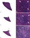
Three-month-old male Sprague Dawley rat treated with 1 mg/kg bodyweight of dexamethasone; tissues were collected three hours later. (a) and (b) are thymus and spleen from a concurrent control animal. Using EH, the lesions in (c) would be diagnosed as thymus, cortex: increased apoptosis, increased tingible body macrophages (arrows), and decreased lymphocytes. For (d), the splenic periarteriolar lymphoid sheath would be the compartment specified and similar diagnoses would be noted. Appropriate laboratory-specific severity grades would be used. Hematoxylin and eosin.
Figure 3
Figure 3
Spleens from a Sprague Dawley rat study with more subtle immunotoxic effects to illustrate the use of enhanced histopathology. The spleen from a concurrent control (a and b) shows a densely cellular periarteriolar lymphoid sheath (PALS) region and adjacent follicle (arrows). The spleen from a low dose animal (c and d) would be diagnosed as PALS: lymphocyte decrease, minimal. The follicle in (d; arrow) is densely cellular and comparable with concurrent control (b). The spleen from the high dose animal (e and f) would be diagnosed as PALS: lymphocyte decrease, moderate. A follicle is not present in (f) for comparison. Hematoxylin and eosin.
Figure 4
Figure 4
Thymus glands from the same study as Figure 3. Compared to the control spleen (a and b) the high dose animals (c and d) had up to a 40% decrease in thymus weight, the thymuses appeared smaller at low magnification, and the cortical area appeared smaller. However, the tissue changes were very subtle, described as decreased area and cellularity of cortex (compare d to b). The cortex stained less intensely, a result of decreased cellularity. This lesion would be difficult to identify without careful comparison to control tissue. Hematoxylin and eosin.
Figure 5
Figure 5
Thymus cortex from two 3-month-old control Sprague Dawley rats within the same study. This is an example of how sectioning and/or staining artifact can interfere with enhanced histopathology evaluation. The thymus in (a) was sectioned thicker and thus appeared more cellular than the thymus in (b). The ideal section thickness is 5 µm. Without knowledge of tissue thickness differences, the tissue in (a) might be misdiagnosed as increased cellularity or the tissue in (b) might be misdiagnosed as decreased lymphocyte cellularity. Hematoxylin and eosin.

Similar articles

Cited by

References

    1. Basketter DA, Bremmer JN, Buckley P, Kammuller ME, Kawabata T, Kimber I, Loveless SE, Magda S, Stringer DA, Vohr HW. Pathology considerations for, and subsequent risk assessment of, chemicals identified as immunosuppressive in routine toxicology. Food Chem Toxicol. 1995;33:239–243. - PubMed
    1. Cesta MF. Normal structure, function, and histology of mucosaassociated lymphoid tissue. Toxicol Pathol. 2006a;34:599–608. - PubMed
    1. Cesta MF. Normal structure, function, and histology of the spleen. Toxicol Pathol. 2006b;34:455–465. - PubMed
    1. Elmore SA. Enhanced histopathology of the lymph nodes. Toxicol Pathol. 2006a;34:634–647. - PMC - PubMed
    1. Elmore SA. Enhanced histopathology of the spleen. Toxicol Pathol. 2006b;34:648–655. - PMC - PubMed

Publication types