. 2012 Feb;86(3):1661-9.
doi: 10.1128/JVI.06073-11. Epub 2011 Nov 16.

Xpr1 is an atypical G-protein-coupled receptor that mediates xenotropic and polytropic murine retrovirus neurotoxicity

Affiliations

Xpr1 is an atypical G-protein-coupled receptor that mediates xenotropic and polytropic murine retrovirus neurotoxicity

Andrew E Vaughan et al. J Virol. 2012 Feb.

Abstract

Xenotropic murine leukemia virus-related virus (XMRV) was first identified in human prostate cancer tissue and was later found in a high percentage of humans with chronic fatigue syndrome (CFS). While exploring potential disease mechanisms, we found that XMRV infection induced apoptosis in SY5Y human neuroblastoma cells, suggesting a mechanism for the neuromuscular pathology seen in CFS. Several lines of evidence show that the cell entry receptor for XMRV, Xpr1, mediates this effect, and chemical cross-linking studies show that Xpr1 is associated with the Gβ subunit of the G-protein heterotrimer. The activation of adenylate cyclase rescued the cells from XMRV toxicity, indicating that toxicity resulted from reduced G-protein-mediated cyclic AMP (cAMP) signaling. Some proteins with similarity to Xpr1 are involved in phosphate uptake into cells, but we found no role of Xpr1 in phosphate uptake or its regulation. Our results indicate that Xpr1 is a novel, atypical G-protein-coupled receptor (GPCR) and that xenotropic or polytropic retrovirus binding can disrupt the cAMP-mediated signaling function of Xpr1, leading to the apoptosis of infected cells. We show that this pathway is also responsible for the classic toxicity of the polytropic mink cell focus-forming (MCF) retrovirus in mink cells. Although it now seems clear that the detection of XMRV in humans was the result of sample contamination with a recombinant mouse virus, our findings may have relevance to neurologic disease induced by MCF retroviruses in mice.


Figures
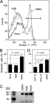
Fig 1
Fig 1
XMRV induces apoptosis in SY5Y cells. (A) Representative fluorescence-activated cell sorter (FACS) histogram showing annexin V staining of SY5Y cells 3 days after infection with the indicated retroviruses. The black bar represents the gate used to determine the percentage of apoptotic cells. FITC, fluorescein isothiocyanate. (B) Quantitation of apoptosis measured by FACS analysis 3 days after infection. Results are means ± standard deviations (SD) from 7 experiments. (C) XIAP expression 3 days after retrovirus infection of SY5Y cells was determined by Western blotting using equal amounts of total cell protein (Bradford assay [4a]) and XIAP antibody (catalog number 610762; BD Biosciences). (D) Quantitation of apoptosis by FACS analysis 3 days after exposure of SY5Y cells to medium made by the transfection of the VP62 XMRV plasmid (VP62 TX) or a control plasmid expressing GFP (GFP-TX) or to medium containing XMRV from 22Rv1 cells. Results are means ± SD from 2 experiments.
Fig 2
Fig 2
Expression of mXpr1 protects SY5Y cells from XMRV-induced apoptosis. SY5Y cells transduced with retroviral vectors encoding the indicated Xpr1 proteins or the empty vector were exposed to medium containing the indicated retroviruses or fresh medium only (mock), and the percentages of apoptotic cells were measured 3 days later. Results are means ± SD from two (empty vector and human Xpr1) or three (mouse Xpr1) experiments.
Fig 3
Fig 3
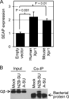
Overexpression of mXpr1 or hXpr1 increases cAMP levels, and hXpr1 is associated with the Gβ G-protein subunit. (A) cAMP levels in HEK 293 cells were measured by using the Cre-SEAP reporter 24 h after transduction with vectors encoding mXpr1 or hXpr1 or with the empty vector, as indicated. Results are means ± SD from three independent experiments. (B) SY5Y cells were incubated with IgG Fc-tagged NZB virus or amphotropic retrovirus Env SU proteins or with no protein (mock). Total proteins were cross-linked, coimmunoprecipitated using anti-IgG Fc antibody, and analyzed for the presence of the Gβ protein by Western blotting with a pan-Gβ antibody. Proteins present in the cell lysate made after protein cross-linking (input) and after coimmunoprecipitation (Co-IP) are shown. Lower bands in the coimmunoprecipitation lanes are due to dissociated bacterial protein G from the Sepharose beads. Input lanes indicate equivalent amounts of Gβ in the cell lysates prior to coimmunoprecipitation.
Fig 4
Fig 4
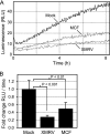
XMRV and MCF 98D virus infection of SY5Y cells decreases intracellular cAMP levels. (A) SY5Y cells containing the Glosensor cAMP 22F reporter construct were infected with virus and assayed for luciferase activity 3 days later. Luminescence as a function of time following the addition of luciferin to the cells is plotted. Each line represents the average of data from three wells in a 96-well plate. RLU, relative light units. (B) Fold changes in rates of luminescence gain relative to those of mock-infected cells are shown. Results are means ± SD from two experiments.
Fig 5
Fig 5
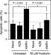
Forskolin protects SY5Y cells from XMRV toxicity. SY5Y cells were cultured with or without 50 μM forskolin for 24 h prior to infection and for 72 h after infection with the indicated viruses. Results are means ± SD from seven experiments. Note that the data for untreated (no forskolin) cells are also shown in Fig. 1B. The statistical analysis depicted here was performed by ANOVA using all data from untreated and treated cells, while that for Fig. 1B used only the data for untreated cells.
Fig 6
Fig 6
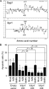
Truncation mutants of Xpr1 provide protection against XMRV-induced apoptosis. (A) Hydropathy plots of the yeast (S. cerevisiae) Syg1 and human Xpr1 proteins are shown. The Xpr1 plot was shifted to the right to align the first putative transmembrane domain of each protein. The locations of the last amino acids of the C-terminal truncation mutations are shown. The 400- and 417-aa Syg1 truncations were found to be fully active in yeast (34). (B) SY5Y cells transduced with retroviral vectors encoding the indicated hXpr1 truncation mutants, or the empty vector, were cultured and infected with the indicated retroviruses. Three days after infection, the percentages of apoptotic cells were measured by annexin V staining and flow cytometry. Results are means ± SD from three independent experiments.
Fig 7
Fig 7
Forskolin protects mink cells from polytropic (MCF) virus-induced death. Twenty-percent-confluent Mv1Lu mink lung epithelial cells (ATCC CCL-64) were exposed to ∼107 infectious units of MCF 98D virus or no virus in the presence of 4 μg/ml Polybrene. Cells were trypsinized and seeded at 1:10 and 1:40 dilutions every time they reached confluence. Cells were visually monitored daily for the presence of any cytopathic effects and cell death. Cells were treated with 25 μM forskolin (FSK) as indicated. Cell numbers were normalized to the starting cell number and were approximated based on the time that it took to reach confluence again after a 1:10 or 1:40 split. Results using two different preparations of MCF virus harvested from MDTF cells infected with the MCF 98D virus were the same.
Fig 8
Fig 8
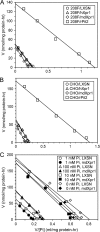
Expression of human or Mus dunni Xpr1 does not result in increased levels of phosphate uptake or significant alterations in the regulation of uptake. (A) Retroviral vectors encoding human Xpr1 (hXpr1) or Mus dunni Xpr1 (mdXpr1) were expressed in 208F rat cells that were used for phosphate uptake analysis. Eadie-Hofstee plots of phosphate uptake velocity (V) versus the velocity/phosphate concentration ratio (V/[Pi]) are shown. The y axis regression line intercept gives the Vmax for phosphate uptake, and the slope is equal to −Km. (B) The same phosphate uptake experiment described above for panel A was performed by using CHO cells. (C) 208F cells transduced with the mdXpr1-expressing vector or the empty vector LXSN were incubated with the indicated concentrations of phosphate overnight, and phosphate uptake was measured.

References

    1. Aloia AL, et al. 2010. XMRV: a new virus in prostate cancer? Cancer Res. 70: 10028–10033 - PMC - PubMed
    1. Battini JL, Rasko JE, Miller AD. 1999. A human cell-surface receptor for xenotropic and polytropic murine leukemia viruses: possible role in G protein-coupled signal transduction. Proc. Natl. Acad. Sci. U. S. A. 96: 1385–1390 - PMC - PubMed
    1. Biedler JL, Roffler-Tarlov S, Schachner M, Freedman LS. 1978. Multiple neurotransmitter synthesis by human neuroblastoma cell lines and clones. Cancer Res. 38: 3751–3757 - PubMed
    1. Bigner DD, et al. 1981. Heterogeneity of genotypic and phenotypic characteristics of fifteen permanent cell lines derived from human gliomas. J. Neuropathol. Exp. Neurol. 40: 201–229 - PubMed
    1. Bradford MM. 1976. A rapid and sensitive method for the quantitation of microgram quantities of protein utilizing the principle of protein-dye binding. Anal. Biochem. 72: 248–254 - PubMed

Publication types

Substances

LinkOut - more resources