. 2012 Mar;26(3):1064-76.
doi: 10.1096/fj.11-196220. Epub 2011 Nov 16.

Schwann cell-specific JAM-C-deficient mice reveal novel expression and functions for JAM-C in peripheral nerves

Affiliations

Schwann cell-specific JAM-C-deficient mice reveal novel expression and functions for JAM-C in peripheral nerves

Bartomeu Colom et al. FASEB J. 2012 Mar.

Abstract

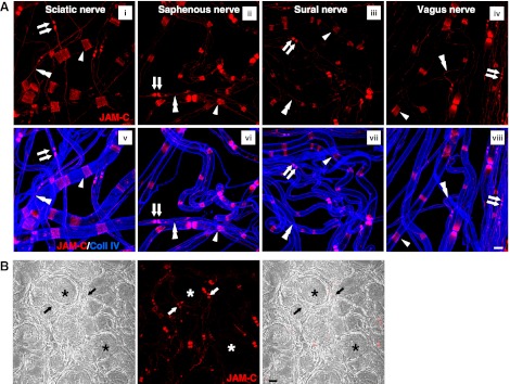
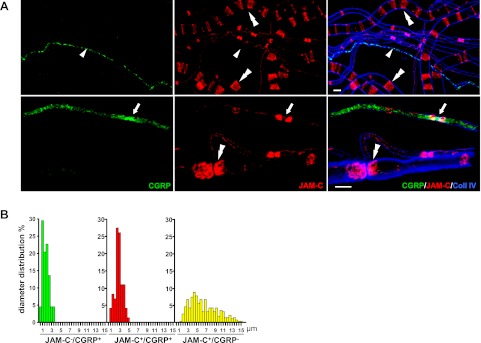
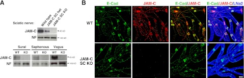
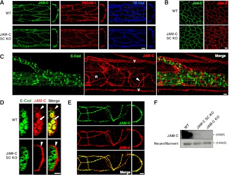
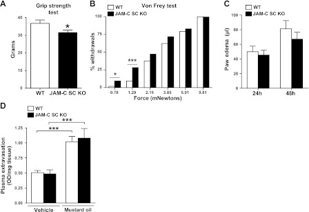
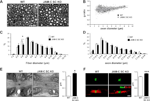
Junctional adhesion molecule-C (JAM-C) is an adhesion molecule expressed at junctions between adjacent endothelial and epithelial cells and implicated in multiple inflammatory and vascular responses. In addition, we recently reported on the expression of JAM-C in Schwann cells (SCs) and its importance for the integrity and function of peripheral nerves. To investigate the role of JAM-C in neuronal functions further, mice with a specific deletion of JAM-C in SCs (JAM-C SC KO) were generated. Compared to wild-type (WT) controls, JAM-C SC KO mice showed electrophysiological defects, muscular weakness, and hypersensitivity to mechanical stimuli. In addressing the underlying cause of these defects, nerves from JAM-C SC KO mice were found to have morphological defects in the paranodal region, exhibiting increased nodal length as compared to WTs. The study also reports on previously undetected expressions of JAM-C, namely on perineural cells, and in line with nociception defects of the JAM-C SC KO animals, on finely myelinated sensory nerve fibers. Collectively, the generation and characterization of JAM-C SC KO mice has provided unequivocal evidence for the involvement of SC JAM-C in the fine organization of peripheral nerves and in modulating multiple neuronal responses.


Figures

Figure 1.
Figure 1.
JAM-C expression in different nerves. A) Confocal images showing immunostaining of JAM-C and collagen IV in whole-mounted teased fibers from sciatic (i, v), saphenous (ii, vi), sural (iii, vii), and vagus (iv, viii) nerves. In all nerves studied, JAM-C was expressed at paranodal regions (arrows) on both side of the nodes of Ranvier, SLIs (arrowheads) and mesaxons (double arrowheads). B) Dorsal root ganglia sections showed lack of JAM-C in the neuronal cell bodies (asterisks), but positive expression was seen in paranodes of the fibers originating directly from them (arrows). Scale bars = 10 μm.
Figure 2.
Figure 2.
JAM-C is expressed in medium- but not small-diameter murine sensory fibers. A) Expression profiles of JAM-C and CGRP discriminate 3 subpopulations of fibers in mouse sciatic nerves: JAM-C/CGRP+, unmyelinated small sensory C fibers (arrowheads); JAM-C+/CGRP+, thinly myelinated Aδ sensory fibers (arrows); and JAM-C+/CGRP, myelinated fibers, including large motor fibers, a subset of Aδ fibers, and Aβ fibers (double arrowheads). B) Diameter distribution of the 3 fiber subpopulations described in panel A (JAM-C/CGRP+, n=44; JAM-C+/CGRP+, n=73; JAM-C+/CGRP, n=286 fibers). Scale bars: 10 μm.
Figure 3.
Figure 3.
Characterization of SC-specific JAM-C-KO mice. A) WB analysis showed deletion of JAM-C as compared to neurofilament-M (NF) in sciatic, sural, saphenous, and vagus nerves from JAM-C SC KO mice as compared to littermate WT or heterozygous controls (5 μg of homogenate protein was loaded). B) Analysis of sciatic nerve teased fibers by confocal microscopy confirmed the deletion of JAM-C from SCs in JAM-C SC KO as compared to WT mice. Expression of E-cadherin and laminin-α2 showed no differences between strains. Scale bar = 10 μm.
Figure 4.
Figure 4.
Expression of JAM-C in endothelial, epithelial, and perineural cells from JAM-C SC KO mice. A) Longitudinal (left panels) and cross-sectional (right panels) confocal images of cremasteric venules showing normal expression of JAM-C at junctions of ECs (stained for PECAM-1 and VE-cadherin) from JAM-C SC KO as compared to WT mice. B) Confocal images of epithelial cells (stained for JAM-A) from JAM-C SC KO as compared to WT mice. C) Images of whole-mount cremaster muscles from JAM-C SC KO mice demonstrated deletion of JAM-C from SC (stained for E-cadherin) in peripheral tissues. Unexpectedly, JAM-C (arrowheads) was found to be expressed surrounding peripheral nerve bundles (n). Specific staining of JAM-C is shown by the positive staining of microvascular ECs (v). D) Cross-section analysis of confocal images from cremasteric nerve bundles showed expression of JAM-C at both perineural (arrowheads) and SCs (arrows) in WT animals, whereas JAM-C SC KO mice only expressed JAM-C in perineural cells (arrowheads). E) Expression of JAM-C and JAM-A at junctions of perineural cells from JAM-C SC KO mice. F) WB of sciatic nerves from WT, JAM-C SC KO, and JAM-C complete-KO mice (50 μg of homogenate protein was loaded). Neurofilament-M was used as a loading control. Scale bars = 10 μm.
Figure 5.
Figure 5.
JAM-C SC KO mice exhibit mild neural functional and behavioral defects. A, B) JAM-C SC KO mice showed reduced grip strength (A) and higher withdrawal response to Von Frey hairs (B) as compared to WT controls (n=9 mice/group), indicating muscular weakness and mechanical hypersensitivity, respectively. C, D) JAM-C SC KO mice showed no significant differences as compared to WT in two models of neurogenic inflammation: 24- and 48-h carrageenan-induced paw edema formation (C; n=8 mice/group) and mustard oil-induced ear skin plasma extravasation (D; n=3–5 animals/group). *P < 0.05; ***P<0.001.
Figure 6.
Figure 6.
Neural morphology analysis of JAM-C SC KO mice. A) Representative images of transverse semithin sections of WT and JAM-C SC KO sciatic nerve fibers. B) Scatter plot of g ratio according to axonal diameter of sciatic nerves from WT and JAM-C SC KO mice. C, D) Distribution of sciatic nerve fiber (C) and axon (D) diameters from WT and JAM-C SC KO mice. E, F) Average length of the node of Ranvier was quantified from WT and JAM-C SC KO mouse sciatic nerves by EM (E; n=19 and 30 nodes, respectively, from 3 mice/group) and by confocal microscopy (F) with teased sciatic nerve fibers immunostained for Nav channel and claudin-19 (n=113 and 109 nodes, respectively, from 3 mice/group). Scale bars = 20 μm (A); 2 μm (E, F). *P < 0.05; ***P < 0.001.

References

    1. Bradfield P. F., Nourshargh S., Aurrand-Lions M., Imhof B. A. (2007) JAM family and related proteins in leukocyte migration. Arterioscl. Thromb. Vasc. Biol. 27, 2104–2112 - PubMed
    1. Weber C., Fraemohs L., Dejana E. (2007) The role of junctional adhesion molecules in vascular inflammation. Nat. Rev. 7, 467–477 - PubMed
    1. Santoso S., Sachs U. J., Kroll H., Linder M., Ruf A., Preissner K. T., Chavakis T. (2002) The junctional adhesion molecule 3 (JAM-3) on human platelets is a counterreceptor for the leukocyte integrin Mac-1. J. Exp. Med. 196, 679–691 - PMC - PubMed
    1. Liang T. W., Chiu H. H., Gurney A., Sidle A., Tumas D. B., Schow P., Foster J., Klassen T., Dennis K., DeMarco R. A., Pham T., Frantz G., Fong S. (2002) Vascular endothelial-junctional adhesion molecule (VE-JAM)/JAM 2 interacts with T, NK, and dendritic cells through JAM 3. J. Immunol. 168, 1618–1626 - PubMed
    1. Lamagna C., Meda P., Mandicourt G., Brown J., Gilbert R. J., Jones E. Y., Kiefer F., Ruga P., Imhof B. A., Aurrand-Lions M. (2005) Dual interaction of JAM-C with JAM-B and alpha(M)beta2 integrin: function in junctional complexes and leukocyte adhesion. Mol. Biol. Cell 16, 4992–5003 - PMC - PubMed

Publication types

MeSH terms

LinkOut - more resources