. 2012 Jan;23(2):233-46.
doi: 10.1091/mbc.E11-07-0650. Epub 2011 Nov 16.

YPR139c/LOA1 encodes a novel lysophosphatidic acid acyltransferase associated with lipid droplets and involved in TAG homeostasis

Affiliations

YPR139c/LOA1 encodes a novel lysophosphatidic acid acyltransferase associated with lipid droplets and involved in TAG homeostasis

Sophie Ayciriex et al. Mol Biol Cell. 2012 Jan.

Abstract

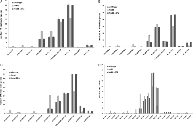
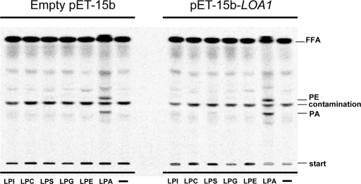
For many years, lipid droplets (LDs) were considered to be an inert store of lipids. However, recent data showed that LDs are dynamic organelles playing an important role in storage and mobilization of neutral lipids. In this paper, we report the characterization of LOA1 (alias VPS66, alias YPR139c), a yeast member of the glycerolipid acyltransferase family. LOA1 mutants show abnormalities in LD morphology. As previously reported, cells lacking LOA1 contain more LDs. Conversely, we showed that overexpression results in fewer LDs. We then compared the lipidome of loa1Δ mutant and wild-type strains. Steady-state metabolic labeling of loa1Δ revealed a significant reduction in triacylglycerol content, while phospholipid (PL) composition remained unchanged. Interestingly, lipidomic analysis indicates that both PLs and glycerolipids are qualitatively affected by the mutation, suggesting that Loa1p is a lysophosphatidic acid acyltransferase (LPA AT) with a preference for oleoyl-CoA. This hypothesis was tested by in vitro assays using both membranes of Escherichia coli cells expressing LOA1 and purified proteins as enzyme sources. Our results from purification of subcellular compartments and proteomic studies show that Loa1p is associated with LD and active in this compartment. Loa1p is therefore a novel LPA AT and plays a role in LD formation.


Figures

FIGURE 1:
FIGURE 1:
Abnormal LDs in loa1Δ and overexpressed LOA1 strains. (A) Wild-type cells and loa1Δ mutant cells were grown in YPD medium until stationary phase. (B) Wild-type strain transformed with the empty expression vector or with the yeast expression vector pYES2/CT-LOA1 were grown on synthetic defined media. Nile red–stained cells were observed under a fluorescence microscope.
FIGURE 2:
FIGURE 2:
Effects of LOA1 on neutral lipid composition. (A) Logarithmic growth phase labeling: steady-state total lipid profiles of wild-type and loa1Δ mutant cells grown until logarithmic phase (OD600 0.5) and labeled with [14C]acetate for an additional 20 h. Lipids were extracted and separated by one-dimensional TLC using first a polar lipid solvent system (midplate) and then a neutral lipid system, as described in Materials and Methods. The results show the amount of [14C]acetate incorporated into individual neutral and polar lipid classes. Dark bar, wild-type cells; gray bar, loa1Δ mutant. Results are mean ± SD from one experiment performed in triplicate and are representative of two distinct experiments. (B) Stationary growth phase labeling: wild-type and loa1Δ mutant cells grown to end of logarithmic phase (OD600 ∼7–8) were labeled with [14C] acetate for 20 h. (B) Incorporation of label into individual lipid classes. Inset, incorporation of label into neutral and polar lipids. Dark bar, wild-type cells; gray bar loa1Δ mutant. Results are mean ± SD from one experiment performed in triplicate and are representative of two distinct experiments. NL, neutral lipids; LP, polar lipids.
FIGURE 3:
FIGURE 3:
Lipid profiling of WT, loa1Δ deletion, and loa1Δ deletion overexpressing LOA1 strains. Cells were grown on synthetic defined media in the presence of 2% galactose and harvested at the early logarithmic phase. Lipids were extracted from yeast cells according to Ejsing et al. (2009). Species were profiled on a LTQ-Orbitrap mass spectrometer in negative mode: (A) PA; or in positive mode: (B) PC, (C) DAG, and (D) TAG. Lipid classes were identified and quantified by the software LipidXplorer (Herzog et al., 2011). Lipid molecular species were determined manually by fragmentation experiments. The abundance of lipid species is depicted in mol%. Error bars indicate ± SD (n = 3 independent analyses).
FIGURE 4:
FIGURE 4:
Acyltransferase activities associated with membranes of E. coli expressing LOA1. Membrane proteins were obtained from C41 (DE3) E. coli transformed with pET-15b (lane 1) and with pET-15b containing the coding sequence of LOA1 (lane 2). Lane 3: negative control without protein; lane 4: positive control with 1 μg yeast protein. LPA AT activities were determined using 0.5 nmol [14C]oleoyl-CoA, 1 nmol 16:0 LPA, and 10 μg membrane-bound proteins as described in Materials and Methods. After a 5-min incubation, lipids were extracted and analyzed by TLC using chloroform/methanol/1-propanol/methyl acetate/0.25% aqueous KCl (10:4:10:10:3.6) as solvent; this was followed by radioimaging. Results are from one experiment that is representative of three experiments performed with independent membrane preparations.
FIGURE 5:
FIGURE 5:
Loa1p exhibits a strict specificity for LPA. Lysophospholipid acyltransferase activities were analyzed using LPI, LPC, LPS, LPG, LPE, and LPA as acyl acceptors (1 nmol). A control without exogenous lysophospholipid (−) was included. Other conditions were as described in Figure 4. Results are from one experiment representative of three experiments performed with independent membrane preparations.
FIGURE 6:
FIGURE 6:
Enzymatic assay of recombinant protein Loa1-V5-6xHis immobilized onto anti-V5 agarose affinity gel antibody. The strain YSA01 (or YSA00 in control) was grown on synthetic defined media supplemented with galactose until the exponential phase. Purification of the recombinant protein was done with anti-V5 agarose affinity gel antibody. After incubation of the yeast lysate and the beads overnight at 4°C, the protein elution was performed with V5 peptide as indicated in Materials and Methods. LPA AT activities were determined using 0.5 nmol [14C]oleoyl-CoA, 1 nmol 16:0 LPA, and several volumes of the fraction of interest. After a 10-min incubation, lipids were extracted and analyzed by TLC using the polar system; this was followed by radioimaging.
FIGURE 7:
FIGURE 7:
Localization of Loa1-HAp in LD. Cells expressing the LOA1-HA fusion gene grown on YPD media until the exponential phase. The tagged gene obtained by homologous recombination was expressed from its own promoter. Cell lysate was loaded on top of a sucrose cushion and centrifuged at 100,000 × g for 90 min. Fractions were collected from the top (T), middle (M), and bottom (B). Proteins were separated by SDS–AGE 12.5% gel and immunoblotted with antisera for the compartment markers: VATPase for the vacuole, Pam1p for the plasma membrane, RFPp for the LDs, Dpm1p for the ER, and Pep12p for the trans-Golgi network/prevacuolar compartment.
FIGURE 8:
FIGURE 8:
Loa1-mCherryp partitioned between ER and large spherical structures, likely LD. Cells expressing the LOA1-mCherry and ERG1-GFP fusion genes grown on YPD media until early log phase (top) or stationary phase (bottom) were examined under fluorescence microscopy as described in Materials and Methods. Merge indicates that fluorescence overlapped; DIC: differential interference contrast. Scale bars: 5 μm.
FIGURE 9:
FIGURE 9:
Localization of Loa1-Ds red1p in LDs. Cells expressing the LOA1-Ds red1 fusion gene grown on YPD media until the exponential phase were stained with BODIPY 493/503 and examined under a fluorescence microscope as described in Materials and Methods. The fusion gene obtained by homologous recombination was expressed with its own promoter. Con A, concanavalin A-treated cell walls. BODIPY shows the LDs. The Ds red1 signal shows the localization of the fusion protein. Merge indicates that fluorescence overlapped.
FIGURE 10:
FIGURE 10:
Purified LD catalyzed PA biosynthesis. Enzymatic activities were determined using 0.5 nmol [14C]oleoyl-CoA, 1 nmol 16:0 LPA, and LD proteins purified from wild-type and loa1Δ deletion strains. After a 10-min incubation, lipids were extracted and analyzed by TLC using the polar system; this was followed by radioimaging.
FIGURE 11:
FIGURE 11:
Loa1p is selective for oleoyl-CoA. LPA AT activities were determined by incubating 2 μg LD proteins purified from the slc1Δ deletion strain with 1 nmol 16:0 LPA and various amounts of [14C]oleoyl-CoA or [14C]palmitoyl-CoA. After a 10-min incubation time, lipids were extracted and analyzed by TLC using the polar system and monitored by radioimaging.

References

    1. Athenstaedt K, Zweytick D, Jandrositz A, Kohlwein SD, Daum G. Identification and characterization of major lipid particle proteins of the yeast Saccharomyces cerevisiae. J Bacteriol. 1999;181:6441–6448. - PMC - PubMed
    1. Athenstaedt K, Daum G. Biosynthesis of phosphatidic acid in lipid particles and endoplasmic reticulum of Saccharomyces cerevisiae. J Bacteriol. 1997;179:7611–7616. - PMC - PubMed
    1. Beaudoin F, Wilkinson BM, Stirling CJ, Napier JA. In vivo targeting of a sunflower oil body protein in yeast secretory (sec) mutants. Plant J. 2000;23:159–70. - PubMed
    1. Benghezal M, Roubaty C, Veepuri V, Knudsen J, Conzelmann A. SLC1 and SLC4 encode partially redundant acyl-coenzyme A 1-acylglycerol-3-phosphate O-acyltransferases of budding yeast. J Biol Chem. 2007;282:30845–30855. - PubMed
    1. Bonangelino CJ, Chavez EM, Bonifacino JS. Genomic screen for vacuolar protein sorting genes in Saccharomyces cerevisiae. Mol Biol Cell. 2002;13:2486–2501. - PMC - PubMed

Publication types

MeSH terms

Substances