. 2012 Jan;23(1):213-24.
doi: 10.1091/mbc.E11-05-0415. Epub 2011 Nov 16.

RNA-related nuclear functions of human Pat1b, the P-body mRNA decay factor

Affiliations

RNA-related nuclear functions of human Pat1b, the P-body mRNA decay factor

Aline Marnef et al. Mol Biol Cell. 2012 Jan.

Abstract

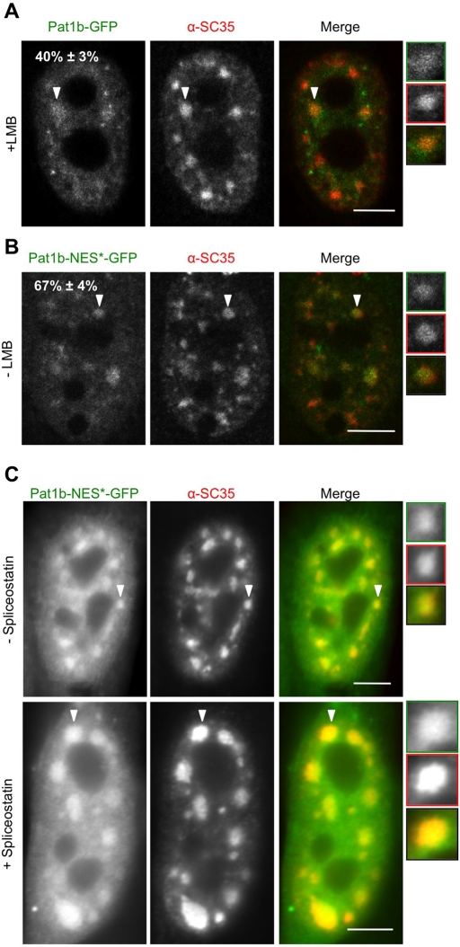
The evolutionarily conserved Pat1 proteins are P-body components recently shown to play important roles in cytoplasmic gene expression control. Using human cell lines, we demonstrate that human Pat1b is a shuttling protein whose nuclear export is mediated via a consensus NES sequence and Crm1, as evidenced by leptomycin B (LMB) treatment. However, not all P-body components are nucleocytoplasmic proteins; rck/p54, Dcp1a, Edc3, Ge-1, and Xrn1 are insensitive to LMB and remain cytoplasmic in its presence. Nuclear Pat1b localizes to PML-associated foci and SC35-containing splicing speckles in a transcription-dependent manner, whereas in the absence of RNA synthesis, Pat1b redistributes to crescent-shaped nucleolar caps. Furthermore, inhibition of splicing by spliceostatin A leads to the reorganization of SC35 speckles, which is closely mirrored by Pat1b, indicating that it may also be involved in splicing processes. Of interest, Pat1b retention in these three nuclear compartments is mediated via distinct regions of the protein. Examination of the nuclear distribution of 4E-T(ransporter), an additional P-body nucleocytoplasmic protein, revealed that 4E-T colocalizes with Pat1b in PML-associated foci but not in nucleolar caps. Taken together, our findings strongly suggest that Pat1b participates in several RNA-related nuclear processes in addition to its multiple regulatory roles in the cytoplasm.


Figures

FIGURE 1:
FIGURE 1:
Crm1-dependent nuclear accumulation of Pat1b but not Pat1a. Fluorescence imaging of HeLa cells treated with LMB for 5 h (+LMB) or with methanol as a control (−LMB). Cells were also stained with DAPI. Scale bar, 10 μm. (A) Localization of endogenous Pat1b using a α-Pat1b antibody. (B, C) Cells were transfected with GFP-tagged Pat1b (B) or Pat1a (C) and treated 24 h later with LMB. Pat1b-GFP accumulates to the nucleus upon LMB treatment (B), whereas Pat1a-GFP localization remains unchanged (C). Note that the nuclear signal seen with the Pat1b antibody in untreated cells is due to an unrelated cross-reacting protein, as it is unresponsive to Pat1b siRNA (Marnef et al., 2010).
FIGURE 2:
FIGURE 2:
Identification of the Pat1b NES sequence. (A) Schematic representation of the localization of different Pat1b truncations tagged with GFP; C and N, cytoplasmic and nuclear localization respectively. The boundaries between the various regions (I–V) are annotated with their corresponding amino acid residues. The NES sequence is highlighted in red, and in green when mutated. (B, C) Fluorescence imaging of HeLa cells treated with LMB for 5 h (+LMB) or with methanol as a control (−LMB). Cells were also stained with DAPI. Scale bar, 10 μm. (B) Shuttling of GFP-tagged Pat1b-N-terminal half (Pat1b-Nter-GFP) upon LMB treatment. (C) Mutation in Pat1b NES (Pat1b-NES*-GFP) causes it to accumulate to the nucleus in the absence of LMB. Four point mutations in Pat1b-NES*-GFP promote its retention to the nucleus in untreated cells. (D) Alignment of six vertebrate Pat1b protein putative NES sequences. Mm1b (Mus musculus Pat1b), Rn1b (Rattus norvegicus Pat1b), Cf1b (Canis familiaris Pat1b), Hs1b (Homo sapiens Pat1b), Xl1b (X. laevis Pat1b), and Dr1b (Danio renio Pat1b). The red amino acids indicate those mutated in human Pat1b NES. Stars indicate conserved hydrophobic residues.
FIGURE 3:
FIGURE 3:
Most P-body components are not nucleocytoplasmic proteins. (A) Fluorescence imaging of HeLa cells treated with LMB for 5 h (+LMB) or with methanol as a control (−LMB) stained with antibodies against P-body components as indicated, including 4E-T, Dcp1a, rck/p54, Edc3, Ge-1, and Xrn1. Scale bar, 10 μm. (B) Graph of % of cells containing indicated numbers of P-bodies in the presence (+) or absence (−) of LMB, as seen with α-rck/p54.
FIGURE 4:
FIGURE 4:
Pat1b localizes to nuclear speckles. Confocal imaging of HeLa cells treated with LMB for 5 h (+LMB) or with methanol as a control (−LMB) stained with the speckle marker SC35 (red). Scale bar, 5 μm. (A) Pat1b-GFP significantly colocalizes to nuclear speckles in the presence of LMB in 40 ± 3% of cells. (B) Pat1b-NES*GFP mutant also colocalizes to speckles, as seen with SC35, in the absence of LMB in 67 ± 4% of cells. (A, B) White arrowheads point to speckles zoomed in the box. (C) Fluorescence imaging of HeLa cells transfected with Pat1b-NES*-GFP and treated with 200 nM spliceostatin A for 5 h (+spliceostatin), or with methanol as a control (−spliceostatin) and stained with SC35 antibodies (red). Scale bars, 5 μm.
FIGURE 5:
FIGURE 5:
Pat1b-GFP localizes to nuclear foci in LMB-treated cells, which associate with PML bodies but not telomeres, centromeres, or heterochromatin. (A–D) Fluorescence imaging of HeLa cells (A, C, and D) or U2OS cells (B) treated with LMB for 5 h (+LMB). Scale bar, 5 μm. (A) Pat1b-GFP localizes to nuclear foci in 25 ± 2% of cells, which were stained with DAPI. (B, C) Pat1b-GFP does not colocalize with telomeres, as seen with α-TRF1 antibody (B), or with centromeres, as seen with α-CENP-A antibody (C). (D) HP1-GFP was cotransfected with Pat1b-HA visualized with α-HA antibody, and Pat1b foci do not colocalize with heterochromatin sites. (E, F) Confocal imaging of HeLa cells treated with LMB for 5 h (+LMB) and stained with a monoclonal α-PML antibody. (E) Pat1b-GFP nuclear foci are adjacent to PML bodies, as seen in example 1, or they overlap with PML bodies, as seen in example 2. (F) Pat1b-GFP can also colocalize with PML bodies, as seen in examples 1 and 2.
FIGURE 6:
FIGURE 6:
Endogenous Pat1b is associated with PML bodies in FOG cells. (A, B) Fluorescence imaging and (C) confocal imaging of FOG cells costained with α-Pat1b (red) and α-PML antibodies (green). Scale bar, 10 μm. (A) Cells were treated with (+LMB) or without LMB (−LMB). (B) Left, si-Pat1b-transfected (+siRNA) and control cells (−siRNA) were compared in the presence of LMB. The zoomed box contains an example of a nucleus containing Pat1b-foci. Right, Western blot of UT (untransfected), si-Pat1b or control si-β-globin, probed with α-Pat1b and α-S6K as a loading control. (C) Pat1b nuclear foci are adjacent (top right, zoomed box) or coalescent (bottom left, zoomed box) to PML bodies.
FIGURE 7:
FIGURE 7:
Pat1b-GFP nuclear foci colocalize with 4E-T foci in LMB-treated cells. HeLa cells treated with LMB for 5 h. (A) Fluorescence imaging of 4E-T-GFP localizing to nuclear foci; nucleus was stained with DAPI. (B) Confocal imaging of cells cotransfected with HA-tagged Pat1b, visualized using an α-HA antibody, and GFP-tagged 4E-T. Most 4E-T nuclear foci colocalize with Pat1b foci, as seen in example 1. However a few foci do not colocalize, as shown in examples 2 and 3. (C) Fluorescence imaging of 4E-T-GFP showing a relationship with PML bodies, identified with a monoclonal PML antibody. Most 4E-T nuclear foci are adjacent to PML bodies, as seen in example 1, some overlap (example 2), and some colocalize (example 3). Scale bar, 5 μm.
FIGURE 8:
FIGURE 8:
Pat1b N-ter mediates its localization to nuclear foci, whereas its C-ter promotes its localization to nuclear speckles. (A, B) Fluorescence imaging of HeLa cells treated with LMB for 5 h. Scale bar, 5 μm. (A) Pat1b N-ter-GFP localizes to nuclear foci in 17 ± 1% cells. These foci may associate with PML bodies, as seen with a monoclonal PML antibody. (B) Pat1b-C-ter displays nuclear speckles in 59 ± 5% of cells, as visualized with antibody to SC35, a speckle marker. (C) Expression levels of GFP constructs transfected in HeLa cells in the presence (+LMB) or absence of LMB (−LMB). Total cell extracts of GFP-tagged Pat1b N-ter and C-ter analyzed by Western blot, using an α-GFP antibody and α-actin antibody (as a loading control).
FIGURE 9:
FIGURE 9:
Inhibition of transcription leads to the loss of nuclear foci and the redistribution of Pat1b to the nucleolus. Fluorescence imaging of HeLa cells treated simultaneously with Act D and LMB for 5 h (A, C) or with Act D only (B) and stained with DAPI and a α-SC35 antibody. Scale bar, 5 μm. (A) Pat1b-GFP displays a diffuse nucleoplasmic localization in 64 ± 8% of cells treated with both LMB and Act D, with a partial relocalization of Pat1b-GFP to a part of the nucleolus, to nucleolar caps (white arrow and zoomed box). (B) Pat1b-NES*-GFP localizes to the nucleolar cap in 42 ± 6% of cells only treated with Act D. (C) Pat1b-Nter-GFP relocalizes to nucleolar caps in 88 ± 3% of cells, whereas Pat1b-Cter-GFP does not (D). (E) 4E-T-GFP displays a nucleocytoplasmic localization in 96 ± 2% of cells treated with both LMB and Act D.

Similar articles

Cited by

References

    1. Aizer A, Brody Y, Ler LW, Sonenberg N, Singer RH, Shav-Tal Y. The dynamics of mammalian P body transport, assembly, and disassembly in vivo. Mol Biol Cell. 2008;19:4154–4166. - PMC - PubMed
    1. Amaro AC, Samora CP, Holtackers R, Wang E, Kingston IJ, Alonso M, Lampson M, McAinsh AD, Meraldi P. Molecular control of kinetochore-microtubule dynamics and chromosome oscillations. Nat Cell Biol. 2010;12:319–329. - PMC - PubMed
    1. Andrei MA, Ingelfinger D, Heintzmann R, Achsel T, Rivera-Pomar R, Lührmann R. A role for eIF4E and eIF4E-transporter in targeting mRNPs to mammalian processing bodies. RNA. 2005;11:717–727. - PMC - PubMed
    1. Bhattacharyya S, Sandy A, Groden J. Unwinding protein complexes in ALTernative telomere maintenance. J Cell Biochem. 2010;109:7–15. - PMC - PubMed
    1. Bhattacharyya SN, Habermacher R, Martine U, Closs EI, Filipowicz W. Relief of microRNA-mediated translational repression in human cells subjected to stress. Cell. 2006;125:1111–1124. - PubMed

Publication types

MeSH terms