. 2011 Nov 16;31(46):16517-28.
doi: 10.1523/JNEUROSCI.1787-11.2011.

Disrupting effect of drug-induced reward on spatial but not cue-guided learning: implication of the striatal protein kinase A/cAMP response element-binding protein pathway

Affiliations

Disrupting effect of drug-induced reward on spatial but not cue-guided learning: implication of the striatal protein kinase A/cAMP response element-binding protein pathway

Mathieu Baudonnat et al. J Neurosci. .

Abstract

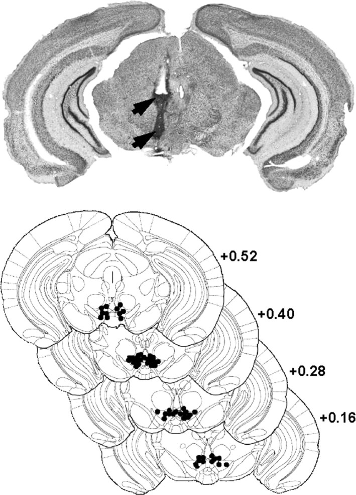
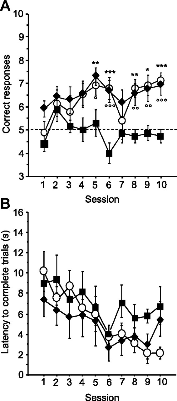
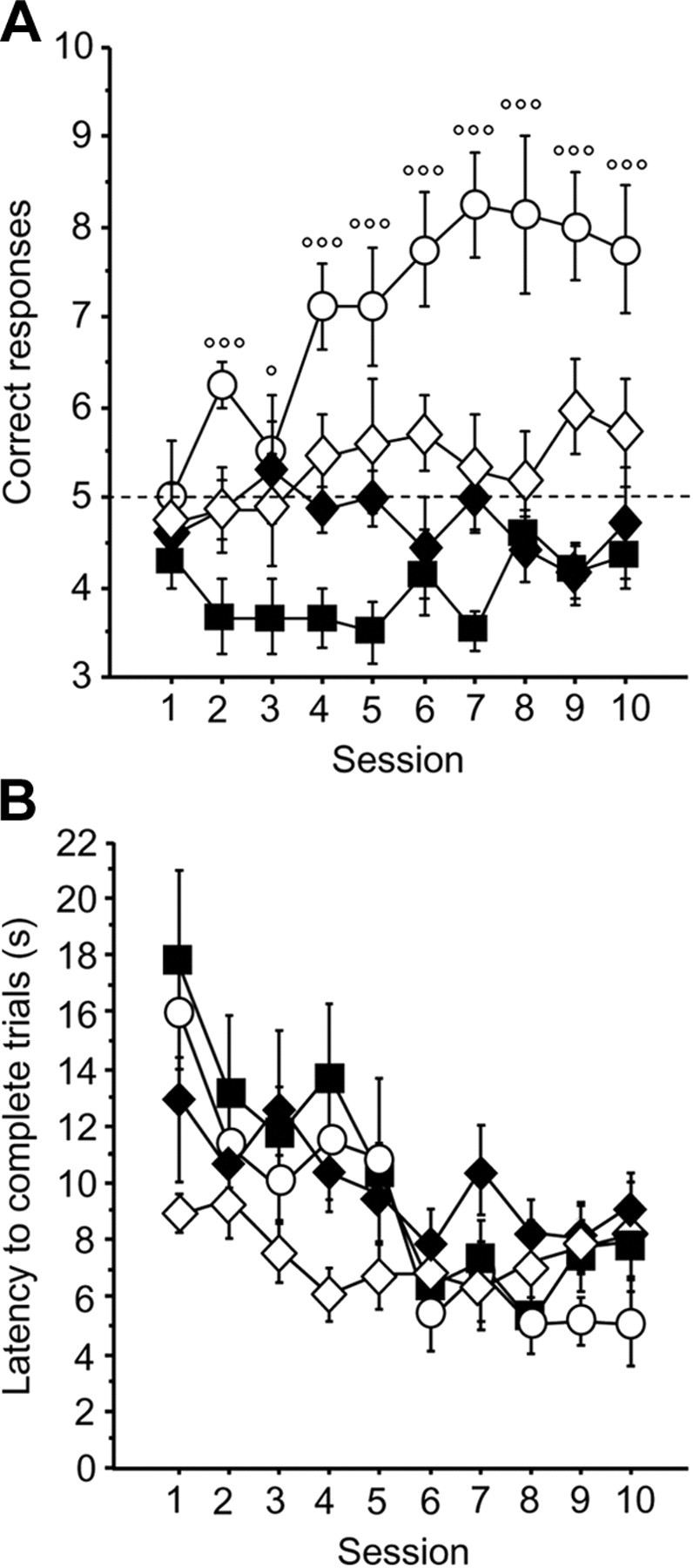
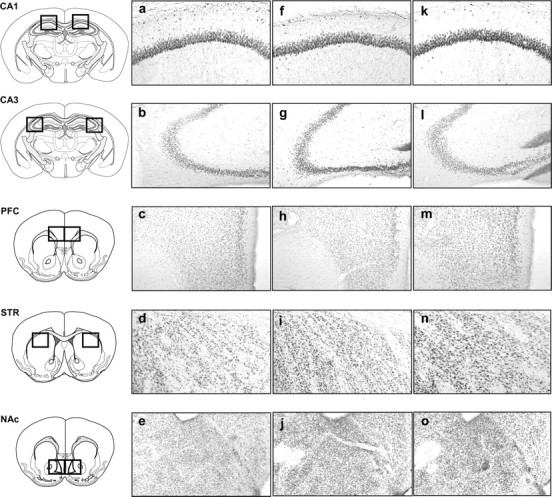
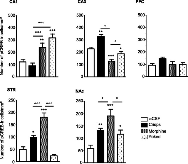
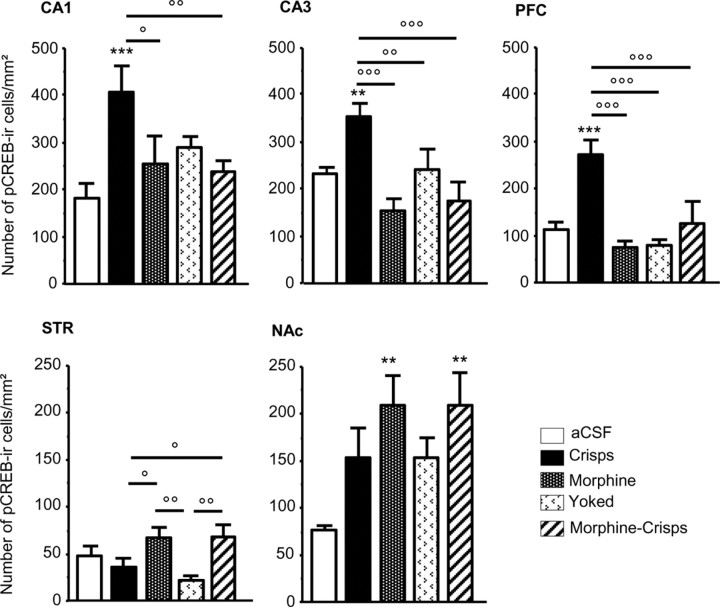
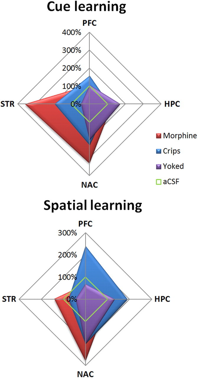
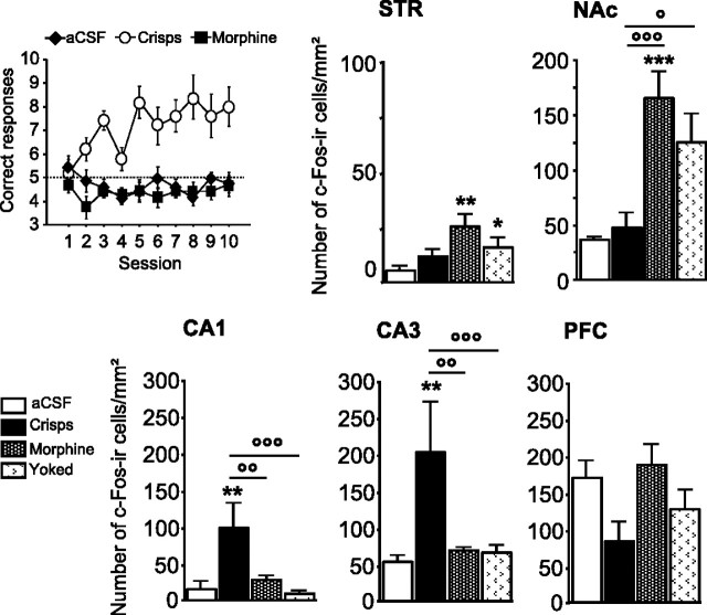
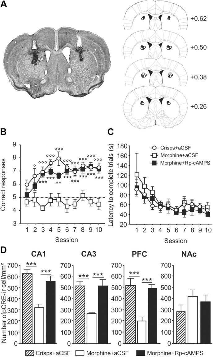
The multiple memory systems hypothesis posits that different neural circuits function in parallel and may compete for information processing and storage. For example, instrumental conditioning would depend on the striatum, whereas spatial memory may be mediated by a circuit centered on the hippocampus. However, the nature of the task itself is not sufficient to select durably one system over the other. In this study, we investigated the effects of natural and pharmacological rewards on the selection of a particular memory system during learning. We compared the effects of food- or drug-induced activation of the reward system on cue-guided versus spatial learning using a Y-maze discrimination task. Drug-induced reward severely impaired the acquisition of a spatial discrimination task but spared the cued version of the task. Immunohistochemical analysis of the phosphorylated form of the cAMP response element binding (CREB) protein and c-Fos expression induced by behavioral testing revealed that the spatial deficit was associated with a decrease of both markers within the hippocampus and the prefrontal cortex. In contrast, drug reward potentiated the cued learning-induced CREB phosphorylation within the dorsal striatum. Administration of the protein kinase A inhibitor 8-Bromo-adenosine-3',5'-cyclic monophosphorothioate Rp isomer (Rp-cAMPS) into the dorsal striatum before training completely reversed the drug-induced spatial deficit and restored CREB phosphorylation levels within the hippocampus and the prefrontal cortex. Therefore, drug-induced striatal hyperactivity may underlie the declarative memory deficit reported here. This mechanism could represent an important early step toward the development of addictive behaviors by promoting conditioning to the detriment of more flexible forms of memory.


Figures

Figure 1.
Figure 1.
Localization of injection sites in the VTA. Top, Wide representative microphotographic view of one injection site, showing traces left by the guide cannula (top arrow) and injection cannula (bottom arrow). Bottom, Histological control of all stereotaxically implanted mice. Black dots show locations of the tip of the cannula (stereotaxic coordinates: anteroposterior, +0.4 mm from interaural; mediolateral, ±0.3 mm; dorsoventral, +3.3 mm). Distribution of self-injection sites corresponds mainly to the posteromedial VTA projection system (Ikemoto, 2007).
Figure 2.
Figure 2.
Acquisition of the cue-guided Y-maze protocol expressed as the mean ± SEM number of correct responses over 10 training sessions (10 trials/d). A, Both natural (crisps group, white dots) and pharmacological (morphine group, black diamonds) rewards allowed the acquisition of this task compared with aCSF (black squares) control group (aCSF vs crisps group: *p < 0.05, **p < 0.01, ***p < 0.001; aCSF vs morphine group: °p < 0.05, °°p < 0.01, °°°p < 0.001). B, Analysis of mean ± SEM latencies to complete a trial (in seconds) over the 10 training sessions (session effect, p < 0.001).
Figure 3.
Figure 3.
Acquisition of the spatial Y-maze protocol expressed as mean ± SEM correct responses over 10 training sessions (10 trials/d). A, Mice reinforced with crisps (white dots) rapidly learned to locate the rewarded arm, exhibiting more correct choices than the three other groups (p < 0.001; crisps vs morphine: °p < 0.05, °°°p < 0.001) (morphine group, black diamonds; morphine–crisps group, white diamonds; aCSF group, black squares). B, Analysis of mean ± SEM latency to complete a trial (in seconds) over the 10 training sessions in the Y-maze (session effect, p < 0.001).
Figure 4.
Figure 4.
Representative photomicrographs of pCREB immunopositive neurons within the CA1 subfield of the hippocampus (CA1), the CA3 subfield of the hippocampus (CA3), the prelimbic part of the PFC, the dorsal STR, and the NAc consequently to the cue-guided protocol: a–e, aCSF group; f–j, crisps group; k–o, morphine group (×10 magnification).
Figure 5.
Figure 5.
Region-specific patterns of CREB phosphorylation following acquisition of the cue-guided learning task. Measures are expressed as mean ± SEM number of pCREB immunoreactive (pCREB-ir) cells per square millimeter in the CA1, CA3, PFC, STR, and NAc. Comparison with aCSF group: *p < 0.05, **p < 0.01, ***p < 0.001; other comparisons: °p < 0.05, °°°p < 0.001.
Figure 6.
Figure 6.
Representative photomicrographs of pCREB immunopositive neurons in the CA1, CA3, PFC, STR, and NAc consequently to the spatial task: a–e, aCSF group; f–j, crisps group; k–o, morphine group (×10 magnification).
Figure 7.
Figure 7.
Region-specific patterns of CREB phosphorylation following acquisition of the spatial learning task. Measures are expressed as mean ± SEM number of pCREB immunoreactive (pCREB-ir) cells per square millimeter in the CA1, CA3, PFC, STR, and NAc. Comparison with aCSF group: **p < 0.01, ***p < 0.001; other comparisons: °p < 0.05, °°p < 0.01, °°°p < 0.001.
Figure 8.
Figure 8.
Summary of pCREB immunostaining changes relative to aCSF animals (100%). Food-reinforced animals exhibited a task-dependent pattern of expression, with a strong activation of hippocampus (HPC) and PFC after spatial learning, whereas the cue-guided task elicited pCREB within the NAc and dorsal STR. This dissociation pattern was absent in drug-reinforced mice, which displayed greater activations of both ventral and dorsal STR after either the spatial or the cue-guided task.
Figure 9.
Figure 9.
Learning-induced brain regional changes in c-Fos immunoreactivity after 10 training days in the spatial version of the Y-maze arm discrimination task. Top left graph shows learning performances in subjects used for the c-Fos study. Measures are expressed as mean ± SEM number of c-Fos immunoreactive (c-Fos-ir) cells per square millimeter in the STR, NAc, CA1, CA3, and PFC. Comparison with aCSF group: **p < 0.01, ***p < 0.001; other comparisons: °p < 0.05, °°p < 0.01, °°°p < 0.001.
Figure 10.
Figure 10.
Acquisition of the spatial version of the Y-maze arm discrimination task in morphine-rewarded animals infused with either Rp-cAMPS or aCSF into the dorsal STR before each training session. A, Schematic representation of injection sites in the dorsal STR. Left, Wide representative microphotographic view of injection sites, showing tracks of guide cannulas (white track) and injection cannulas (black track). Right, Histological control of all stereotaxically implanted mice. Dots show locations of the tip of injection cannulas (black dots, Rp-cAMPS injection; white dots, aCSF injection) (stereotaxic coordinates: anteroposterior, 0.62 mm to bregma; lateral, ±1.9 mm; ventral, 1.5 mm). B, Morphine-rewarded mice pretreated with Rp-cAMPS exhibited more correct responses per session compared with aCSF-injected animals (p < 0.001; morphine + Rp-cAMPS vs morphine + aCSF: **p < 0.01, ***p < 0.001). As expected, crisps-rewarded animals made more correct responses than morphine-rewarded mice when both groups were preinfused with aCSF into the dorsal STR (p < 0.001; crisps + aCSF vs morphine + aCSF: °p < 0.05, °°°p < 0.001). C, The treatment did not alter locomotion, as assessed by the time to complete trials, which decreased in all groups over the course of training sessions (p < 0.001). D, Region-specific pattern of pCREB expression after acquisition of the spatial discrimination task. Measures are expressed as mean ± SEM number of pCREB-immunoreactive (pCREB-ir) cells per square millimeter in the CA1, CA3, PFC, and NAc (comparisons with morphine–aCSF-treated group: ***p < 0.001).

Similar articles

Cited by

References

    1. Akirav I, Richter-Levin G. Biphasic modulation of hippocampal plasticity by behavioral stress and basolateral amygdala stimulation in the rat. J Neurosci. 1999;19:10530–10535. - PMC - PubMed
    1. Arnsten AF, Ramos BP, Birnbaum SG, Taylor JR. Protein kinase A as a therapeutic target for memory disorders: rationale and challenges. Trends Mol Med. 2005;11:121–128. - PubMed
    1. Baldwin AE, Sadeghian K, Holahan MR, Kelley AE. Appetitive instrumental learning is impaired by inhibition of cAMP-dependent protein kinase within the nucleus accumbens. Neurobiol Learn Mem. 2002;77:44–62. - PubMed
    1. Balleine BW, Liljeholm M, Ostlund SB. The integrative function of the basal ganglia in instrumental conditioning. Behav Brain Res. 2009;199:43–52. - PubMed
    1. Barrot M, Olivier JD, Perrotti LI, DiLeone RJ, Berton O, Eisch AJ, Impey S, Storm DR, Neve RL, Yin JC, Zachariou V, Nestler EJ. CREB activity in the nucleus accumbens shell controls gating of behavioral responses to emotional stimuli. Proc Natl Acad Sci U S A. 2002;99:11435–11440. - PMC - PubMed

MeSH terms

LinkOut - more resources