

Multimodal quantitative magnetic resonance imaging of thalamic development and aging across the human lifespan: implications to neurodegeneration in multiple sclerosis
- PMID: 22090508
- PMCID: PMC3230860
- DOI: 10.1523/JNEUROSCI.4184-11.2011
Multimodal quantitative magnetic resonance imaging of thalamic development and aging across the human lifespan: implications to neurodegeneration in multiple sclerosis
Abstract
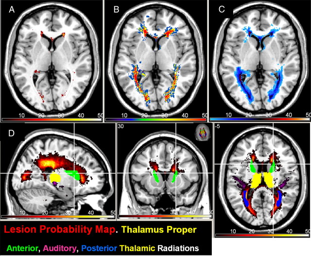
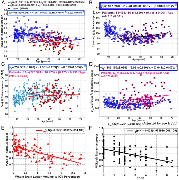
The human brain thalami play essential roles in integrating cognitive, sensory, and motor functions. In multiple sclerosis (MS), quantitative magnetic resonance imaging (qMRI) measurements of the thalami provide important biomarkers of disease progression, but late development and aging confound the interpretation of data collected from patients over a wide age range. Thalamic tissue volume loss due to natural aging and its interplay with lesion-driven pathology has not been investigated previously. In this work, we used standardized thalamic volumetry combined with diffusion tensor imaging, T2 relaxometry, and lesion mapping on large cohorts of controls (N = 255, age range = 6.2-69.1 years) and MS patients (N = 109, age range = 20.8-68.5 years) to demonstrate early age- and lesion-independent thalamic neurodegeneration.
Figures
References
-
- Antulov R, Carone DA, Bruce J, Yella V, Dwyer MG, Tjoa CW, Benedict RH, Zivadinov R. Regionally distinct white matter lesions do not contribute to regional gray matter atrophy in patients with multiple sclerosis. J Neuroimaging. 2011;21:210–218. - PubMed
-
- Aubert-Broche B, Fonov V, Ghassemi R, Narayanan S, Arnold DL, Banwell B, Sled JG, Collins DL. Regional brain atrophy in children with multiple sclerosis. Neuroimage. 2011;58:409–415. - PubMed
-
- Beaulieu C. The basis of anisotropic water diffusion in the nervous system—a technical review. NMR Biomed. 2002;15:435–455. - PubMed
-
- Behrens TE, Johansen-Berg H, Woolrich MW, Smith SM, Wheeler-Kingshott CA, Boulby PA, Barker GJ, Sillery EL, Sheehan K, Ciccarelli O, Thompson AJ, Brady JM, Matthews PM. Non-invasive mapping of connections between human thalamus and cortex using diffusion imaging. Nat Neurosci. 2003;6:750–757. - PubMed
Publication types
MeSH terms
Grants and funding
LinkOut - more resources
Full Text Sources
Medical