Value of ultrasonography-guided fine needle aspiration cytology in the investigative sequence of hepatic lesions with an emphasis on hepatocellular carcinoma
- PMID: 22090691
- PMCID: PMC3214462
- DOI: 10.4103/0970-9371.86344
Value of ultrasonography-guided fine needle aspiration cytology in the investigative sequence of hepatic lesions with an emphasis on hepatocellular carcinoma
Abstract
Background: The evaluation and management of various hepatic lesions is a common clinical problem and their appropriate clinical management depends on accurate diagnoses.
Aims: To study the cytomorphological features of distinctive non-neoplastic and neoplastic lesions of the liver and to evaluate the sensitivity, specificity and diagnostic accuracy of ultrasonography (USG)-guided fine needle aspiration cytology (FNAC) in the diagnosis of liver diseases.
Materials and methods: Seventy-two patients with evidence of liver diseases underwent USG-guided, percutaneous FNAC. Cytomorphological diagnoses were correlated with clinical, biochemical and radiological findings, histopathological diagnoses and follow-up information.
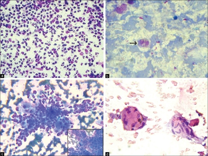
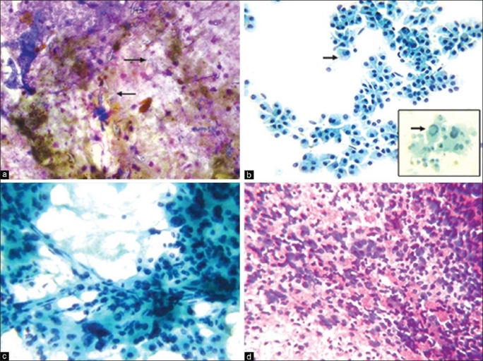
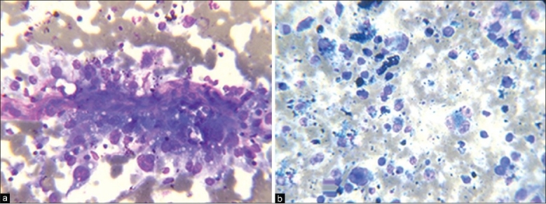
Results: The age of the patients ranged from eight months to 90 years with 48 males (66.67%) and 24 females (33.33%). Of the 72 cases, the cytological diagnosis was rendered in 71 patients and smears were inadequate for interpretation in one case. Neoplastic lesions (68.06%) were more common than non-neoplastic lesions (30.56%). The majority of the neoplastic lesions were hepatocellular carcinomas (36.12%) followed by metastatic adenocarcinomas (19.45%). Among non-neoplastic lesions, cirrhosis was the commonest lesion (8.34%). The overall diagnostic accuracy of FNAC was 97.82% with a sensitivity and specificity of 96.87 and 100% respectively.
Conclusion: USG-guided FNAC of the liver is a safe, simple, cost-effective and accurate method for cytological diagnosis of hepatic diffuse, focal/nodular and cystic lesions with good sensitivity and specificity.
Keywords: Fine needle aspiration cytology; hepatic lesions; ultrasonography.
Conflict of interest statement
Figures
References
-
- Kuo FY, Chen WJ, Lu SN, Wang JH, Eng HL. Fine needle aspiration cytodiagnosis of liver tumors. Acta Cytol. 2004;48:142–8. - PubMed
-
- Leiman G. Liver and Spleen. In: Orell SR, Sterret GF, Whitaker D, editors. Fine needle aspiration cytology. 4th ed. New Delhi: Churchill Livingstone; 2005. pp. 293–316.
-
- Das DK, Tripathi RP, Chachra KL, Sodhani P, Parkash S, Bhambhani S. Role of guided fine needle aspiration cytology in diagnosis and classification of liver malignancies. Trop Gastroenterol. 1997;18:101–6. - PubMed
-
- Tsui WM, Cheng F, Lee Y. Fine needle aspiration cytology of liver tumors. Ann Contemp Diagn Pathol. 1998;2:79–93.
-
- Rasania A, Pandey CL, Joshi N. Evaluation of FNAC in diagnosis of hepatic lesion. J Cytol. 2007;24:51–4.