Pyogenic granuloma associated with periodontal abscess and bone loss - A rare case report
- PMID: 22090773
- PMCID: PMC3214537
- DOI: 10.4103/0976-237X.86478
Pyogenic granuloma associated with periodontal abscess and bone loss - A rare case report
Abstract
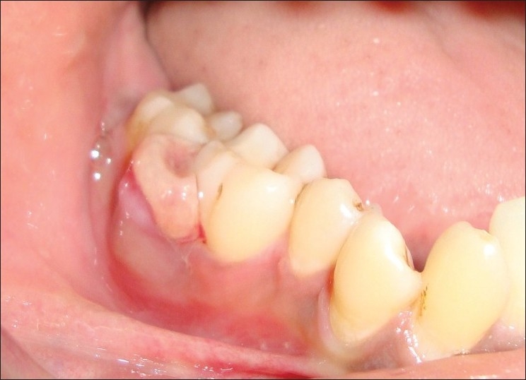
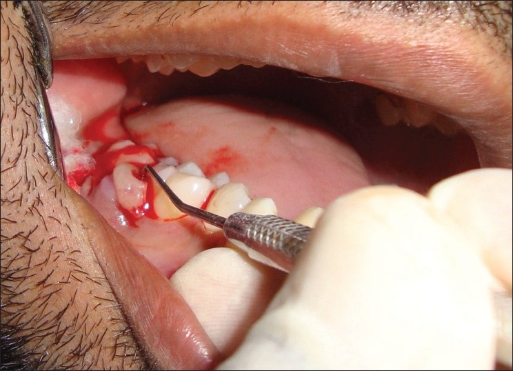
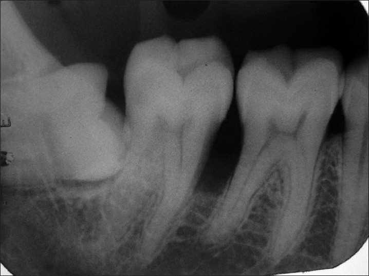
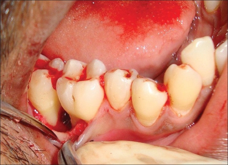
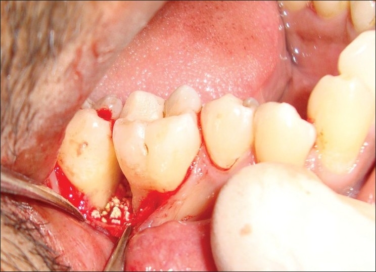
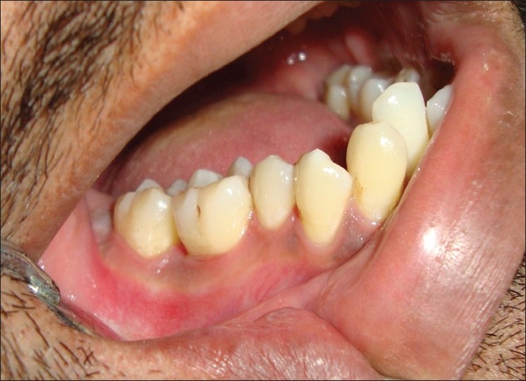
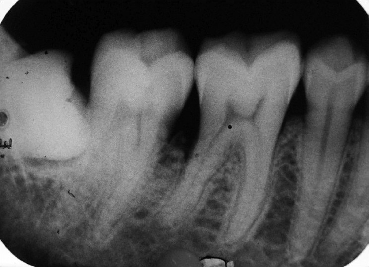
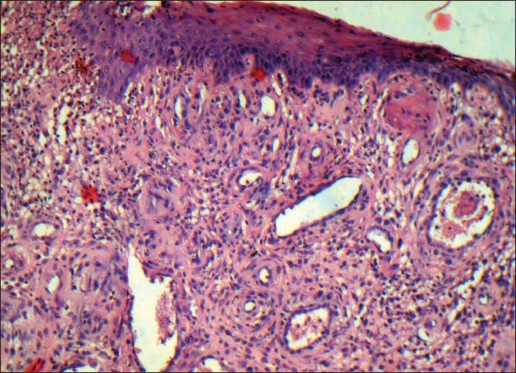
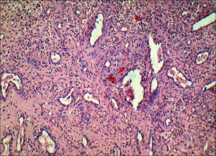
A diverse group of the pathologic process can produce the enlargement of soft tissues in the oral cavity and often present a diagnostic challenge. This soft tissue enlargement may represent a variation of the normal anatomic structure, inflammatory reaction, cyst, neoplasm, and developmental anomalies. A group of reactive hyperplasias, which develop in response to chronic recurring tissue injury that stimulates an excessive tissue repair response. The pyogenic granuloma (PG) is a reactive enlargement that is an inflammatory response to local irritation such as calculus, a fractured tooth, rough dental restoration, and foreign materials or hormonal (pregnancy tumor) and rarely associated with bone loss. This paper presents a rare case of PG associated with periodontal abscess and bone loss in a 30-year-old male.
Keywords: Bone loss; periodontal abscess; pyogenic granuloma.
Conflict of interest statement
Figures
References
-
- Laskaris G. 4th ed. New York: Thieme publishers; 1997. Colour atlas of oral diseases; pp. 400–1.
-
- Shafer WG, Hine MK, Levy BM. 4th ed. Philadelphia: WB Saunders; 1983. A textbook of oral pathology; pp. 359–60.
-
- Daley TD, Wysocki GP, Wysocki PD, Wysocki DM. The major epulides: clinicopathological correlations. J Can Dent Assoc. 1990;56:627–30. - PubMed
-
- Neville BW, Damm DD, Allen CM, Bouquot JE. 2nd ed. Philadelphia: WB Saunders; 2002. Oral and Maxillofacial Pathology; pp. 473–95.
-
- Regezi JA, Sciubba JJ, Jordan RC. 4th ed. Philadelphia: WB Saunders; 2003. Oral pathology: clinical pathologic considerations; pp. 115–6.