. 2011 Sep-Oct;19(5):622-32.
doi: 10.1111/j.1524-475X.2011.00725.x.

Spatiotemporal progression of cell death in the zone of ischemia surrounding burns

Affiliations

Spatiotemporal progression of cell death in the zone of ischemia surrounding burns

Steven T Lanier et al. Wound Repair Regen. 2011 Sep-Oct.

Abstract

Burns are dynamic injuries, characterized by progressive death of surrounding tissue over time. Although central to an understanding of burn injury progression, the spatiotemporal degrees and rates of cellular necrosis and apoptosis in the zone of ischemia surrounding burns are not well characterized. Using a validated porcine hot comb model, we probed periburn tissue at 1, 4, and 24 hours after injury for high-mobility group box 1 as a marker of necrosis and activated cleaved caspase-3 as a marker of apoptosis, followed by spatiotemporal morphometric analysis. We found that necrosis was the most prominent mechanism of cell death in burn injury progression, with significant progression between 1 and 4 hours postburn. Apoptosis appeared not to play a role in early burn injury progression but was observed in cells at the interface of necrotic and viable tissue at 24 hours postburn. Our findings imply that intervention within the first 4 hours following injury is likely necessary to limit burn injury progression. Additionally, based on high-mobility group box 1 staining patterns, we define distinct early, intermediate, and late pathological signs of cell necrosis that may facilitate delineation of causal mechanistic relationships of burn injury progression in vivo.


Figures

Figure 1
Figure 1
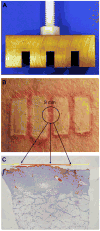
A) Burns are created with a 150 g brass comb with 4 10 × 25 mm prongs separated by 5 mm interspaces. B) Appearance of burn wound immediate after application of the hot comb. 8 mm punch biopsies encompass the 5 mm interspace (zone of ischemia) with 1.5 mm of burned tissue on either side. C) Slide-mounted tissue sample illustrating orientation on slide relative to biopsy placement. For each specimen, a single burn was chosen for analysis with its corresponding zone of ischemia. Boxes display 0.25 mm wide × 1.0 mm deep zones used to quantify endothelial cell necrosis.
Figure 2
Figure 2
HMGB1 controls. A) Unburned dermis stained for HMGB1. The arrow indicates a vascular endothelial cell displaying nuclear HMGB1 staining indicative of cell viability. B) Unburned epithelium stained for HMGB1. Keratinocytes display nuclear HMGB1 staining indicative of cell viability.
Figure 3
Figure 3
Vascular endothelial cell staining of sequential sections. A-C) Tissue from the zone of ischemia stained for Laminin, CD31, and HMGB1, respectively. Notice that necrotic vascular endothelial cell nuclei displayed in Panel C have lost nuclear HMGB1 staining, in comparison to viable endothelial cells shown in Fig. 2A.
Figure 4
Figure 4
Patterns of epidermal HMGB1 staining. A) Absence, B) Diffuse extracellular staining, C) Concentrated extracellular and cytoplasmic staining, D) Nuclear staining without extracellular staining.
Figure 5
Figure 5
HMGB1 staining of burn and interspace at 1, 4, and 24 hours post-burn. Individual panels depict 0.5 mm increments moving from the burn into the zone of ischemia to the right. A) Directly below the hot comb at 1 hour, B-E) 0.5 mm intervals moving away from the burn at 1 hour, F) Directly below the hot comb at 4 hours, G-J) 0.5 mm intervals moving away from the burn at 4 hours, K) Directly below the hot comb at 24 hours, L-O) 0.5 mm intervals moving away from the burn at 24 hours. Arrows mark the progression of epidermal necrosis over time as indicated by loss of keratinocyte nuclear HMGB1 staining. Boxes mark high-powered fields displayed in Figure 6.
Figure 6
Figure 6
Vascular endothelial cell HMGB1 staining at 1, 4, and 24 hours post-burn. A-C) 1 hour post-burn: high-powered views of Fig. 5B-D, respectively. D-F) 4 hours post-burn: High-powered views of Fig. 5G-I, respectively. G-I) 24 hours post-burn: High-powered views of Fig. 5L-N, respectively. Arrows mark vascular endothelial cells with loss of nuclear HMGB1 staining and leakage of HMGB1 into the extracellular space indicative of necrosis.
Figure 7
Figure 7
Spatiotemporal variation in endothelial cell necrosis. A statistically significant increase in percent endothelial necrosis was observed among the 1, 4, and 24-hour post-burn time points (mean ± SE, n = 3 for each time point, P < 0.001).
Figure 8
Figure 8
CC3a staining at 1, 4, and 24 hours post-burn. A) Unburned control, B) 1 hour, C) 4 hours, D) 24 hours. A band of apoptotic cells was observed deep to the area of necrosis, at the interface of necrotic and viable tissue. Panels display the degree of apoptosis for a burn and adjacent 2.5 mm zone of ischemia that spans one half of the entire interspace.

References

    1. Jackson DM. The diagnosis of the depth of burning. Br J Surg. 1953;40(164):588–596. - PubMed
    1. Singh V, Devgan L, Bhat S, Milner SM. The pathogenesis of burn wound conversion. Ann Plast Surg. 2007;59(1):109–15. - PubMed
    1. Gravante G, Delogu D, Palmieri MB, Santeusanio G, Montone A, Esposito G. Inverse relationship between the apoptotic rate and the time elapsed from thermal injuries in deep partial thickness burns. Burns. 2008;34(2):228–33. - PubMed
    1. Gravante G, Palmieri MB, Esposito G, Delogu D, Santeusanio G, Filingeri V, Montone A. Apoptotic death in deep partial thickness burns vs. normal skin of burned patients. J Surg Res. 2007;141(2):141–45. - PubMed
    1. McNamara AR, Zamba KD, Sokolich JC, Jaskille AD, Light TD, Griffin MA, Meyerholz DK. Apoptosis is differentially regulated by burn severity and dermal location. J Surg Res. 2010;162(2):258–63. - PubMed

Publication types

MeSH terms