. 2011 Dec;325(1):92-8.
doi: 10.1111/j.1574-6968.2011.02417.x. Epub 2011 Oct 3.

Changes of lipid domains in Bacillus subtilis cells with disrupted cell wall peptidoglycan

Affiliations
Free PMC article

Changes of lipid domains in Bacillus subtilis cells with disrupted cell wall peptidoglycan

Katarína Muchová et al. FEMS Microbiol Lett. 2011 Dec.
Free PMC article

Abstract

The cell wall is responsible for cell integrity and the maintenance of cell shape in bacteria. The Gram-positive bacterial cell wall consists of a thick peptidoglycan layer located on the outside of the cytoplasmic membrane. Bacterial cell membranes, like eukaryotic cell membranes, are known to contain domains of specific lipid and protein composition. Recently, using the membrane-binding fluorescent dye FM4-64, helix-like lipid structures extending along the long axis of the cell and consisting of negatively charged phospholipids were detected in the rod-shaped bacterium Bacillus subtilis. It was also shown that the cardiolipin-specific dye, nonyl acridine orange (NAO), is preferentially distributed at the cell poles and in the septal regions in both Escherichia coli and B. subtilis. These results suggest that phosphatidylglycerol is the principal component of the observed spiral domains in B. subtilis. Here, using the fluorescent dyes FM4-64 and NAO, we examined whether these lipid domains are linked to the presence of cell wall peptidoglycan. We show that in protoplasted cells, devoid of the peptidoglycan layer, helix-like lipid structures are not preserved. Specific lipid domains are also missing in cells depleted of MurG, an enzyme involved in peptidoglycan synthesis, indicating a link between lipid domain formation and peptidoglycan synthesis.


Figures

Fig. 1
Fig. 1
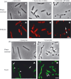
Staining of Bacillus subtilis PY79 cells with fluorescent vancomycin. Fluorescent vancomycin was prepared by mixing BodipyFL vancomycin (Molecular Probes) and unlabeled vancomycin in a 1 : 1 ratio. The mixture was added to the growing cultures to give a final concentration of 1 μg mL−1, and the culture was incubated for a further 20 min. (a) Fluorescent vancomycin staining of vegetatively growing cells. A helical pattern of the fluorophore was observed indicating a helical distribution of the peptidoglycan biosynthetic machinery. Phase-contrast (A) and fluorescence images (B). (b) Fluorescent vancomycin staining of cells treated with lysozyme. Staining was performed as described above. Cells were collected by centrifugation, resuspended in 1× SMM containing 1 mg mL−1 lysozyme, and further incubated for 10 min. The vancomycin fluorescence signal is localized around the circumference of the spheroplasts indicating the presence of residual peptidoglycan. There is no helical pattern to the observed fluorescence in these cells. Phase-contrast (A) and fluorescence images (B). Scale bars represent 2 μm.
Fig. 2
Fig. 2
Lipid domains in spheroplasts of the wild-type Bacillus subtilis strain PY79. (a) Visualization of FM4-64 signals in spheroplasts. Spheroplasts were prepared by enzymatic removal of peptidoglycan with lysozyme. FM4-64-stained anionic phospholipids in wild-type cells resuspended in SMM buffer that were not treated with lysozyme (A) and in cells treated with lysozyme (B and C). (B) Cells with partially removed peptidoglycan after 2 min of lysozyme treatment, and (C) spheroplasts after 10 min of lysozyme treatment. While in the cells untreated with lysozyme, helix-like lipid structures stained with FM4-64 are visible, in spheroplasts, the FM4-64 signals are on the opposite sides of the cells. The arrows show the accumulation of anionic lipids stained with FM4-64. (b) Staining of spheroplasts with NAO to visualize cardiolipin domains. NAO-stained cardiolipin domains in wild-type cells that were not treated with lysozyme (A) and in cells treated with lysozyme (B). NAO fluorescence signals in spheroplasts preponderate at the former cell poles (arrows), indicating enrichment in cardiolipin at these sites as observed in vegetative cells. In cells not treated with lysozyme, arrows show its accumulation at the cell poles and arrowheads show its accumulation at the division sites. Scale bars represent 2 μm.
Fig. 3
Fig. 3
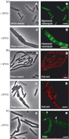
Peptidoglycan staining and localization of lipid domains in IB1302 cells depleted of MurG. Cell cultures were initially grown in the presence of 1 mM IPTG for 2 h before dilution to an OD600 nm of 0.05 in media lacking IPTG and incubation for an additional 2 h. (a) Fluorescent vancomycin staining of peptidoglycan. In cells grown in the presence of IPTG, a helical pattern of fluorescence signal is observed, indicating a helical distribution of the peptidoglycan biosynthetic machinery similar to that in wild-type cells. However, in bulged cells that were grown without IPTG, no clear helical pattern of the fluorophore is observed. Instead, the fluorescence signal is accumulated at division sites where residual peptidoglycan synthesis occurred. Phase-contrast (A) and fluorescence images (B). (b) FM 4-64-stained anionic phospholipids. In the cells grown in the presence of IPTG, lipid domains similar to those observed in wild-type cells were detected. In cells, depleted of MurG, no such lipid structures are observable. Phase-contrast (A) and fluorescence images (B). (c) Cardiolipin domains stained with NAO. The arrow shows cardiolipin accumulation at the cell pole, and the arrowhead shows its accumulation at the division site in a way similar to that seen in wild-type cells. In bulged cells, the fluorescence signal is more evenly distributed in the membrane. Phase-contrast (A) and fluorescence images (B). Scale bars represent 2 μm.

References

    1. Backman K, Ptashne M, Gilbert AW. Construction of plasmids carrying the cI gene of bacteriophage lambda. P Natl Acad Sci USA. 1976;73:4174–4178. - PMC - PubMed
    1. Barák I, Muchová K, Wilkinson AJ, O'Toole PJ, Pavlendová N. Lipid spirals in Bacillus subtilis and their role in cell division. Mol Microbiol. 2008;68:1315–1327. - PMC - PubMed
    1. Bhavsar AP, Brown ED. Cell wall assembly in Bacillus subtilis: how spirals and spaces challenge paradigms. Mol Microbiol. 2006;60:1077–1090. - PubMed
    1. van den Brink-van der Laan E, Boots J-WP, Spelbrink REJ, Kool GM, Breukink E, Killian JA, de Kruijff B. Membrane interaction of the glycosyltransferase MurG: a special role for cardiolipin. J Bacteriol. 2003;185:3773–3779. - PMC - PubMed
    1. Campo N, Tjalsma H, Buist G, et al. Subcellular sites for bacterial protein export. Mol Microbiol. 2004;53:1583–1599. - PubMed

Publication types

LinkOut - more resources