Review
. 2011:81:85-164.
doi: 10.1016/B978-0-12-385885-6.00009-2.

Coronavirus pathogenesis

Affiliations
Review

Coronavirus pathogenesis

Susan R Weiss et al. Adv Virus Res. 2011.

Abstract

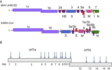
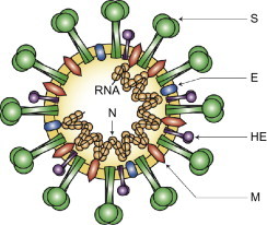
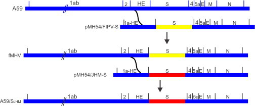
Coronaviruses infect many species of animals including humans, causing acute and chronic diseases. This review focuses primarily on the pathogenesis of murine coronavirus mouse hepatitis virus (MHV) and severe acute respiratory coronavirus (SARS-CoV). MHV is a collection of strains, which provide models systems for the study of viral tropism and pathogenesis in several organs systems, including the central nervous system, the liver, and the lung, and has been cited as providing one of the few animal models for the study of chronic demyelinating diseases such as multiple sclerosis. SARS-CoV emerged in the human population in China in 2002, causing a worldwide epidemic with severe morbidity and high mortality rates, particularly in older individuals. We review the pathogenesis of both viruses and the several reverse genetics systems that made much of these studies possible. We also review the functions of coronavirus proteins, structural, enzymatic, and accessory, with an emphasis on roles in pathogenesis. Structural proteins in addition to their roles in virion structure and morphogenesis also contribute significantly to viral spread in vivo and in antagonizing host cell responses. Nonstructural proteins include the small accessory proteins that are not at all conserved between MHV and SARS-CoV and the 16 conserved proteins encoded in the replicase locus, many of which have enzymatic activities in RNA metabolism or protein processing in addition to functions in antagonizing host response.


Figures

Figure 1
Figure 1
Genome organization and replicase encoded nonstructural proteins. (A) The genomes of MHV-JHM.SD and SARS-CoV are diagrammed. L, leader; ORF1a/1b, replicase; structural genes: HE, hemagglutinin-esterase; S, spike; E, small membrane envelope; M, membrane; N, nucleocapsid; I, internal. orfs encoding accessory genes are designated with numbers. (B). Arrows indicate cleavage sites for orf1a, orf1ab encoded polypeptides and numbers indicate individual nsp cleavage products.
Figure 2
Figure 2
Coronavirus virion structure. The genome RNA is complexed with the N protein to form a helical cased within the viral membrane, HE, hemagglutinin-esterase; S, spike; E, small membrane envelope; M, membrane are all transmembrane proteins.
Figure 3
Figure 3
Targeted recombination. A schematic representation of targeted recombination. A59 sequences are shown in blue, FIPV S ectodomain sequences are shown in yellow, and JHM sequences are shown in red. Based on Kuo et al. (2000).
Figure 4
Figure 4
Assembly strategy of full-length coronavirus genomes. The use of the type II S restriction enzyme Esp3I to ligate two cDNAs of arbitrary sequence. The Esp3I recognition site is shown in underlined upper case text. The arbitrary sequence at which the two cDNAs are joined is shown in lower case text. Based on Yount et al. (2002).
Figure 5
Figure 5
Structures of the JHM.SD and SARS-CoV spike glycoproteins. RBD, receptor-binding domain; HVR, hypervariable region; HR, heptad repeat domain; TM, transmembrane domain. Arrow indicates cleavage site yielding S1 and S2 subunits in JHM.SD spike. Mutations/deletions found in other neurotropic MHV strains and SARS-CoV variants are indicated below structures and discussed in the text.
Figure 6
Figure 6
A speculative evolutionary history of the lectin activity of HE and S proteins. S proteins that maintain the ability to hemaggultinate and bind to glycans are denoted S+; those that have lost this ability as S−. For SARS-CoV, hemagglutinating activity has not been reported and to our knowledge has not been tested for; this is denoted as S? HE proteins with a requirement or a strong preference for 9-O-acetyl sialic acid as their ligand (and esterase substrate) are depicted as HE+. HE proteins from strains of MHV where the preferred ligand (substrate) of HE is 4-O-acetyl sialic acid are depicted as HE + *. This schema is supported by the presence of hemagglutinin (lectin-like) activity of TGEV (an alphacoronavirus) (Krempl et al., 2000) and IBV (a gammacoronavirus) (Niesters et al., 1987), and the presence of a galectin fold in the receptor-binding domain of MHV (Peng et al., 2011).
Figure 7
Figure 7
A schematic diagram of the domain structure of MHV nsp3. The domains depicted are: ubiquitin-related domains (UB1 and UB2), papain-like proteases (PLP-1 and PLP-2), ADP-ribose 1″-phosphatase (ADRP), metal binding domain (MBD), betacoronavirus-specific nucleic acid binding (NAB) and marker (G2M) domains, a putative metal-binding region containing a zinc finger (ZF), and three subdomains forming part of the Y region (Y1–Y3). Transmembrane domains are depicted by vertical bars. Based on Neuman et al., 2008.

Similar articles

Cited by

References

    1. Almazan F., Gonzalez J.M., Penzes Z., Izeta A., Calvo E., Plana-Duran J., Enjuanes L. Engineering the largest RNA virus genome as an infectious bacterial artificial chromosome. Proc. Natl. Acad. Sci. USA. 2000;97:5516–5521. - PMC - PubMed
    1. Almazan F., Dediego M.L., Galan C., Escors D., Alvarez E., Ortego J., Sola I., Zuniga S., Alonso S., Moreno J.L., Nogales A., Capiscol C. Construction of a severe acute respiratory syndrome coronavirus infectious cDNA clone and a replicon to study coronavirus RNA synthesis. J. Virol. 2006;80:10900–10906. - PMC - PubMed
    1. Almeida M.S., Johnson M.A., Herrmann T., Geralt M., Wuthrich K. Novel beta-barrel fold in the nuclear magnetic resonance structure of the replicase nonstructural protein 1 from the severe acute respiratory syndrome coronavirus. J. Virol. 2007;81:3151–3161. - PMC - PubMed
    1. Alvarez E., DeDiego M.L., Nieto-Torres J.L., Jimenez-Guardeno J.M., Marcos-Villar L., Enjuanes L. The envelope protein of severe acute respiratory syndrome coronavirus interacts with the non-structural protein 3 and is ubiquitinated. Virology. 2010;402:281–291. - PMC - PubMed
    1. An S., Chen C.J., Yu X., Leibowitz J.L., Makino S. Induction of apoptosis in murine coronavirus-infected cultured cells and demonstration of E protein as an apoptosis inducer. J. Virol. 1999;73:7853–7859. - PMC - PubMed

Publication types

MeSH terms

Substances