. 2011 Nov 15;20(5):576-90.
doi: 10.1016/j.ccr.2011.09.009.

Direct signaling between platelets and cancer cells induces an epithelial-mesenchymal-like transition and promotes metastasis

Affiliations

Direct signaling between platelets and cancer cells induces an epithelial-mesenchymal-like transition and promotes metastasis

Myriam Labelle et al. Cancer Cell. .

Abstract

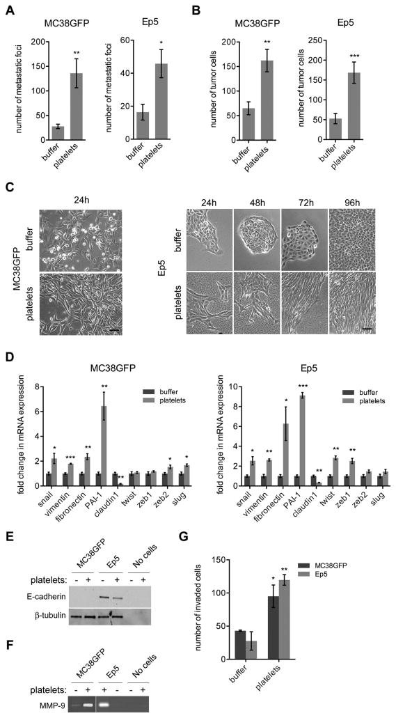
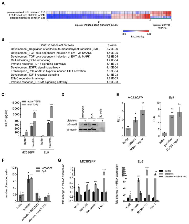
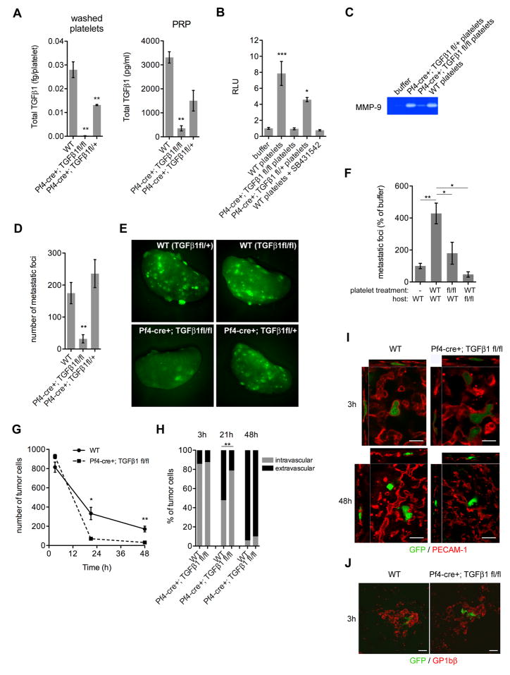
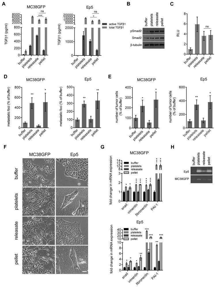
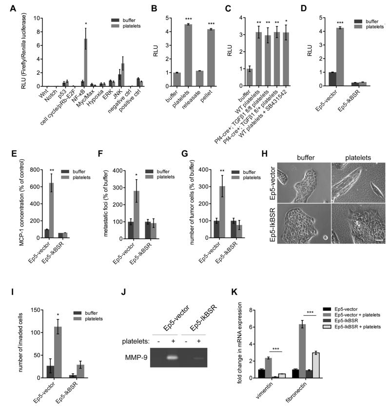
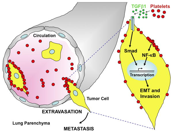
Interactions of cancer cells with the primary tumor microenvironment are important determinants of cancer progression toward metastasis but it is unknown whether additional prometastatic signals are provided during the intravascular transit to the site of metastasis. Here, we show that platelet-tumor cell interactions are sufficient to prime tumor cells for subsequent metastasis. Platelet-derived TGFβ and direct platelet-tumor cell contacts synergistically activate the TGFβ/Smad and NF-κB pathways in cancer cells, resulting in their transition to an invasive mesenchymal-like phenotype and enhanced metastasis in vivo. Inhibition of NF-κB signaling in cancer cells or ablation of TGFβ1 expression solely in platelets protects against lung metastasis in vivo. Thus, cancer cells rely on platelet-derived signals outside of the primary tumor for efficient metastasis.


Figures

Figure 1
Figure 1. Pretreatment of Tumor Cells with Platelets Promotes Lung Metastasis by Increasing Tumor Cell Seeding and Inducing an EMT-Like Invasive Phenotype
(A) Numbers of metastatic foci at the surface of lungs (2 largest lobes) 14 days after tail-vein injection of MC38GFP cells or Ep5 cells stably expressing ZsGreen (Ep5-ZsGreen) pretreated with buffer (vehicle) or platelets for 40h (n=5–12). (B) Numbers of tumor cells at the surface of lungs 48h after tail-vein injection of MC38GFP or Ep5-ZsGreen cells pretreated with buffer or platelets for 40h (n=6–11). (C) Phase-contrast micrographs of MC38GFP or Ep5 cells treated with buffer or platelets for the times indicated. Scale bar=50μm. (D) Relative fold change in mRNA expression in MC38GFP or Ep5 cells treated with buffer or platelets for 40h (n=3). Values are normalized to Gapdh expression. (E) Detection of E-cadherin protein levels by immunoblotting of lysates of MC38GFP or Ep5 cells treated as in (D). Amounts of platelets equal to those used to treat cells were also loaded as control (no cells). β-tubulin was used as loading control. (F) Zymography for MMP-9 in the conditioned medium of MC38GFP or Ep5 cells treated as in (D). Amounts of platelets equal to those used to treat cells were also loaded as control (no cells). (G) MC38GFP and Ep5 cells were added at the top of transwells coated with Matrigel and treated with buffer or platelets. The total number of cells that invaded to the bottom of the transwell was counted after 48h (n=3). For panels A, B, D and G bars represent the mean ± SEM. *p<0.05, **p<0.01, ***p<0.001 were determined by Student’s t-test. See also Figure S1.
Figure 2
Figure 2. Platelet-Induced Gene Expression Signature Reveals Increased Expression of Prometastatic Genes and Activation of the TGFβ Pathway in Tumor Cells
(A) Heat map of genes regulated by more than 4 fold (p<0.05) in Ep5 cells treated with platelets in comparison with untreated Ep5 cells (line 2). Line 1 and line 2 show Log2 ratios of gene expression compared to untreated Ep5 cells. mRNAs present in platelets (line1) were removed from the list of genes modulated upon platelet treatment of Ep5 cells (line 2) to generate a platelet-induced gene signature (line 3), which is listed in Table S2. (B) Canonical signaling pathways most significantly associated with the list of genes differentially expressed by Ep5 cells upon platelet treatment (platelet-induced gene signature; Table S1; threshold = 2 fold, up and down regulated genes considered, p<0.05) as determined with GeneGo canonical pathway maps. The ten pathways with the lowest p values are shown. (C) Concentration of active and total TGFβ1 in conditioned medium from MC38GFP or Ep5 cells treated with buffer or platelets for 40h. The conditioned medium was collected, centrifuged to remove platelets, and the presence of TGFβ1 in the supernatant measured by ELISA. Each bar represents the mean ± SEM of n=2–6. ***p<0.001 as determined by Student’s t-test. (D) Detection of phospho-Smad2 protein levels by immunoblotting of Ep5 or MC38GFP cells treated as in (C). Amounts of platelets equal to those used to treat cells were also loaded as control (no cells). β-tubulin is used as loading control. (E) Relative luciferase activity (RLU) in MC38GFP or Ep5 cells stably expressing a luciferase reporter under the control of an SBE promoter and treated for 40h with buffer, platelets or 1ng/ml TGFβ1 (positive control) (n=5–6). (F) MC38GFP and Ep5 cells were added at the top of transwells coated with Matrigel and treated with buffer, platelets, platelets + SB431542 (10μM) or platelets + TGFβ1 blocking antibody (6μg/ml). The total numbers of cells that invaded to the bottom of the transwell were counted after 48h (n=3). (G) Relative fold change in mRNA expression in MC38GFP or Ep5 cells treated with buffer, or platelets +/− SB431542 (10μM) for 40h (n=3). Values are normalized to Gapdh expression. For panels E, F and G bars represent the mean ± SEM, and *p<0.05, **p<0.01, ***p<0.001 vs buffer were determined by one-way ANOVA followed by Tuckey’s post test.
Figure 3
Figure 3. Platelet-Derived TGFβ Promotes Lung Metastasis
(A) Concentration of TGFβ1 in washed platelets and platelet-rich plasma (PRP) from wild-type (WT), Pf4-cre+; TGFβ1fl/fl or Pf4-cre+; TGFβ1fl/+ mice measured by ELISA (n=2). (B) Relative luciferase activity (RLU) in Ep5 cells stably expressing a luciferase reporter under the control of the SBE promoter and treated with buffer, platelets from wild-type (WT), Pf4-cre+; TGFβ1fl/fl, Pf4-cre+; TGFβ1fl/+, or with wild-type platelets + SB431542 (10μM) for 20h (n=3). (C) Zymography for MMP-9 in the conditioned medium from Ep5 cells treated with buffer, platelets from wild-type (WT), Pf4-cre+; TGFβ1fl/fl or Pf4-cre+; TGFβ1fl/+ mice for 40h. (D–E) Numbers of metastatic foci at the surface of lungs (2 largest lobes) 14 days after tail-vein injection of MC38GFP cells in wild-type (WT), Pf4-cre+; TGFβ1fl/fl or Pf4-cre+; TGFβ1fl/+ mice (n=7–9), and representative pictures of lungs (E). (F) Numbers of metastatic foci at the surface of lungs (2 largest lobes) 14 days after tail-vein injection of MC38GFP cells pretreated with buffer (−), platelets from WT mice (WT) or platelets from Pf4-cre+; TGFβ1fl/fl (fl/fl) and injected into WT or Pf4-cre+; TGFβ1fl/fl mice (n=9–14). (G) Numbers of tumor cells at the surface of lungs 3h, 21h and 48h after tail-vein injection of MC38GFP cells in wild-type (WT) or Pf4-cre+; TGFβ1fl/fl mice. Each point represents the mean ± SEM number of cells/view field (3X) (n=3–14). *p<0.05, **p<0.01 were determined by Student’s t-test. (H) Percentage of intravascular and extravascular MC38GFP cells in lungs of wild-type (WT) or Pf4-cre+; TGFβ1fl/fl mice 3h, 21h and 48h after tail-vein injection of tumor cells (n=16–46 cells). **p<0.01 as determined by Fisher’s exact test. (I–J) Confocal microcopy of lungs of wild-type (WT) or Pf4-cre+; TGFβ1fl/fl mice 3h and 48h after tail-vein injection of tumor cells for MC38GFP cells (green) and either blood vessels (H; PECAM-1 staining; red) or platelets (I; GP1bβ staining; red. Note platelet aggregates surrounding tumor cells.). Scale bar=50μm. For panels A, B, D, and F bars represent the mean ± SEM, and *p<0.05, **p<0.01, ***p<0.001 vs WT or buffer were determined by one-way ANOVA followed by Tuckey’s post test. See also Figure S2 and Table S3.
Figure 4
Figure 4. Platelet-Derived TGFβ1 and Platelet-Bound Factors Cooperate to Promote Metastasis
(A) Concentration of TGFβ measured by ELISA in the conditioned medium of MC38GFP or Ep5 cells incubated with buffer, platelets, releasate from activated platelets (releasate), or the pellet fraction from activated platelets (pellet) for 40h (n=4–6). (B) Detection of phospho-Smad2 and total Smad2 protein levels by immunoblotting in Ep5 cells treated as in (A). β-tubulin was used as loading control. (C) Relative luciferase activity (RLU) in Ep5 cells stably expressing a luciferase reporter under the control of the SBE promoter and treated as in (A) for 20h (n=2). (D) Numbers of metastatic foci at the surface of lungs (2 largest lobes) 14 days after tail-vein injection of MC38GFP or Ep5-ZsGreen cells pretreated with buffer, platelets, releasate from activated platelets (releasate), or the pellet fraction from activated platelets (pellet) for 40h (n=5–17). (E) Numbers of tumor cells at the surface of lungs 48h after tail-vein injection of MC38GFP or Ep5-ZsGreen cells pretreated as in (D). Each bar represents the mean ± SEM number of cells/view field (3X) (n=5–13). *p<0.05, **p<0.01 vs buffer were determined by Student’s t-test. (F) Phase-contrast micrographs of MC38GFP and Ep5 cells treated as in (A) for 24h. Scale bar=50μm. (G) Relative fold change in mRNA expression in MC38GFP or Ep5 cells treated as in (A) for 40h. Values are normalized to Gapdh expression (n=3). (H) Zymography for MMP-9 in the conditioned medium from Ep5 cells treated as in (A) for 40h. For panels A, C, D and G, bars represent the mean ± SEM, and ns (p>0.05), *p<0.05, **p<0.01, ***p<0.001 vs buffer were determined by one-way ANOVA followed by Tuckey’s post test. See also Figure S3 and Table S4.
Figure 5
Figure 5. TGFβ1 and Direct Platelet-Tumor Cell Contact Synergize to Promote Prometastatic Gene Expression
(A) Relative luciferase activity (RLU) in MLEC cells stably expressing a luciferase reporter under the control of a TGFβ responsive PAI-1 promoter construct and treated with different concentrations of TGFβ1 (left panel) or platelets (right panel). Note that the y-axis scale is the same for both panels and that platelets give higher stimulation than achievable with TGFβ1 alone. (B) Relative luciferase activity (RLU) in MLEC cells stably expressing a luciferase reporter under the control of a PAI-1 promoter construct treated with buffer, platelets from WT or Pf4-cre+; TGFβ1fl/fl mice, TGFβ1 1ng/ml or with combinations of platelets + TGFβ1 1ng/ml, +/− SB431542 (SB; 10μM) or +/− TGFβ1 blocking antibody (Ab; 6μg/ml)(n=3–16). (C) Relative luciferase activity (RLU) in MLEC cells stably expressing a luciferase reporter under the control of a PAI-1 promoter construct treated with buffer, platelets, or thrombin-activated platelets +/− 1ng/ml TGFβ1 seeded either at the bottom (direct contact with tumor cells) or in the upper chamber of a transwell (0.4μm pore size) to prevent direct contact between platelets and tumor cells (n=2–3). (D) Zymography for MMP-9 in the conditioned medium from Ep5 or MC38GFP cells treated with buffer, platelets, or thrombin-activated platelets seeded either at the bottom or in the upper chamber of a transwell (0.4μm pore size). (E) Relative fold change in mRNA expression in Ep5 or MC38GFP cells treated as in (D) (n=3). Values are normalized to Gapdh expression. For panels B, C and E, bars represent the mean ± SEM, and *p<0.05, **p<0.01, ***p<0.001 vs buffer (unless otherwise indicated) were determined by one-way ANOVA followed by Tuckey’s post test.
Figure 6
Figure 6. The NF-κB Signaling Pathway is Activated by Platelets in a Contact-Dependent Manner and Cooperates with TGFβ Signaling to Induce an EMT-Like Transition and Promote Metastasis
(A) Ep5 cells were transfected with pathway-specific firefly luciferase reporters and constitutively expressed control Renilla luciferase reporters. 24h after transfection, cells were treated with buffer or platelets for 20h, and the relative luciferase activity (RLU) was measured. Firefly luciferase activity was normalized to Renilla luciferase activity. Each bar represents the mean ± SEM of n=3. *p<0.05 was determined by Student’s t-test. (B) Relative luciferase activity (RLU) in Ep5 cells stably expressing an NF-κB luciferase reporter and treated with buffer, platelets, releasate from activated platelets (releasate), or the pellet fraction from activated platelets (pellet) for 20h (n=2). (C) Relative luciferase activity (RLU) in Ep5 cells stably expressing a NF-κB luciferase reporter and treated with buffer, platelets from wild-type (WT), Pf4-cre+; TGFβ1fl/fl, Pf4-cre+; TGFβ1fl/+ mice or with wild-type platelets + SB431542 (10μM) for 20h (n=5). (D) Relative luciferase activity (RLU) in Ep5 cells stably expressing a NF-κB luciferase reporter and either a IκB super-repressor (Ep5-IkBSR) or a control vector (Ep5-vector) and treated with buffer or platelets for 20h (n=4). (E) MCP-1 concentration in the conditioned medium from Ep5 cells stably expressing an IκB super-repressor (Ep5-IkBSR) or a control vector (Ep5-vector) and treated with buffer or platelets for 20h (n=2). (F) Numbers of metastatic foci at the surface of lungs (2 largest lobes) 14 days after tail-vein injection of Ep5-IkBSR and Ep5-vector cells pretreated with buffer or platelets for 40h. Each bar represents the mean ± SEM of n=5–7. *p<0.05 was determined by Student’s t-test. (G) Numbers of tumor cells at the surface of lungs 48h after tail-vein injection of Ep5-IkBSR and Ep5-vector cells pretreated with buffer or platelets for 40h. Each bar represents the mean ± SEM of n=4–7 **p<0.01 was determined by Student’s t-test. (H) Phase-contrast micrographs of Ep5-IkBSR and Ep5-vector cells treated with buffer or platelets for 24h. Scale bar=50μm. (I) Ep5-IkBSR and Ep5-vector cells were added at the top of transwells coated with Matrigel and treated with buffer or platelets. The total numbers of cells that invaded to the bottom of the transwell were counted after 48h (n=2). (J) Zymography for MMP-9 in the conditioned medium from Ep5-IkBSR and Ep5-vector cells treated with buffer or platelets for 40h. (K) Relative fold change in mRNA expression in Ep5-IkBSR and Ep5-vector cells treated as in (J) (n=3). Values are normalized to Gapdh expression. For panels B, C and K, bars represent the mean ± SEM, and *p<0.05, **p<0.01, ***p<0.001 vs buffer were determined by one-way ANOVA followed by Tuckey’s post test. For panels D, E and I, bars represent the mean ± SEM, and *p<0.05 vs buffer was determined by two-way ANOVA followed by Bonferroni’s post test. See also Figure S4.
Figure 7
Figure 7. Platelet-Tumor Cell Contact and Platelet-Derived TGFβ1 Synergize to Promote an EMT-Like Transition and Metastasis
Platelets secrete TGFβ1, which activates the TGFβ/Smad pathway in tumor cells. Upon direct platelet-tumor cell contact, the NF-κB pathway is also activated in tumor cells and synergizes with TGFβ/Smad signaling to induce a rapid EMT, enhance invasiveness and promote metastasis. Activation of neither the TGFβ/Smad nor the NF-κB pathway alone is sufficient to promote metastasis. Thus, platelet-tumor cell contact triggers a synergistic interaction between TGFβ/Smad and NF-κB pathways that is necessary for efficient metastasis. The metastatic potential of tumor cells therefore continues to evolve outside of the primary tumor site in response to platelet-to-tumor cell signaling.

Comment in

References

    1. Abe M, Harpel JG, Metz CN, Nunes I, Loskutoff DJ, Rifkin DB. An assay for transforming growth factor-beta using cells transfected with a plasminogen activator inhibitor-1 promoter-luciferase construct. Anal Biochem. 1994;216:276–284. - PubMed
    1. Assoian RK, Komoriya A, Meyers CA, Miller DM, Sporn MB. Transforming growth factor-beta in human platelets. Identification of a major storage site, purification, and characterization. J Biol Chem. 1983;258:7155–7160. - PubMed
    1. Assoian RK, Sporn MB. Type beta transforming growth factor in human platelets: release during platelet degranulation and action on vascular smooth muscle cells. J Cell Biol. 1986;102:1217–1223. - PMC - PubMed
    1. Bakewell SJ, Nestor P, Prasad S, Tomasson MH, Dowland N, Mehrotra M, Scarborough R, Kanter J, Abe K, Phillips D, et al. Platelet and osteoclast beta3 integrins are critical for bone metastasis. Proc Natl Acad Sci U S A. 2003;100:14205–14210. - PMC - PubMed
    1. Biswas S, Guix M, Rinehart C, Dugger TC, Chytil A, Moses HL, Freeman ML, Arteaga CL. Inhibition of TGF-beta with neutralizing antibodies prevents radiation-induced acceleration of metastatic cancer progression. J Clin Invest. 2007;117:1305–1313. - PMC - PubMed

Publication types

Substances

Associated data