. 2012 Feb;125(2):418-29.
doi: 10.1093/toxsci/kfr310. Epub 2011 Nov 17.

An NF-κB-independent and Erk1/2-dependent mechanism controls CXCL8/IL-8 responses of airway epithelial cells to cadmium

Affiliations

An NF-κB-independent and Erk1/2-dependent mechanism controls CXCL8/IL-8 responses of airway epithelial cells to cadmium

Estelle Cormet-Boyaka et al. Toxicol Sci. 2012 Feb.

Abstract

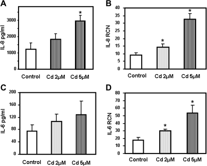
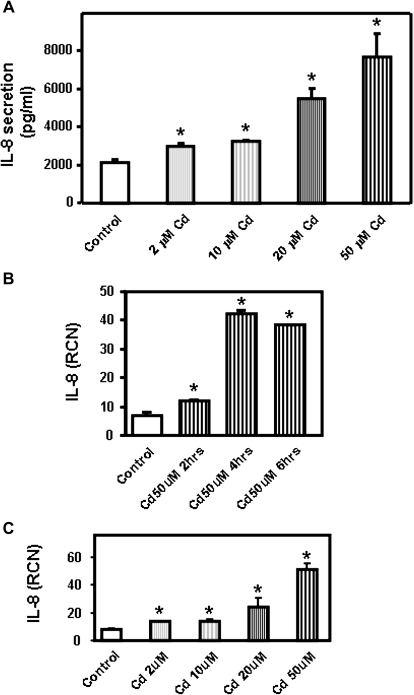
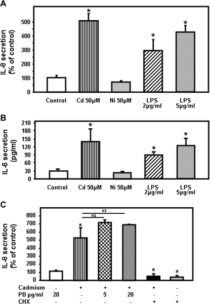
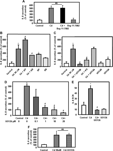
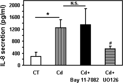
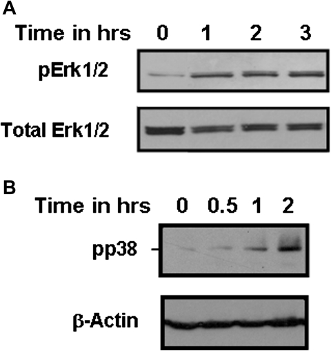
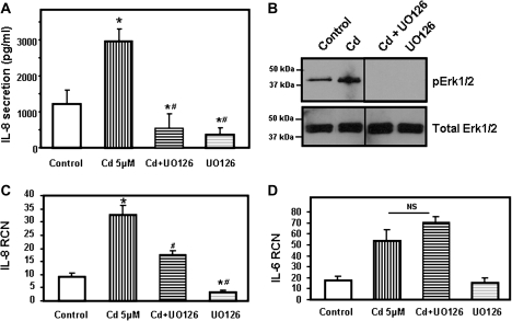
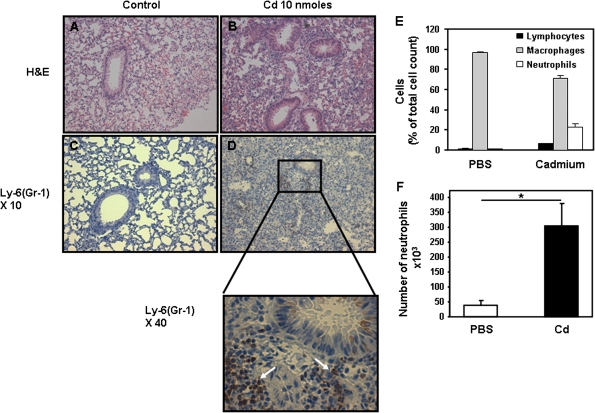
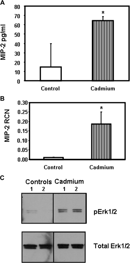
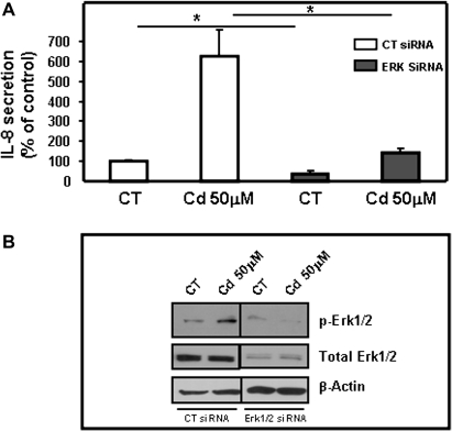
Airway epithelial cells in the lung are the first line of defense against pathogens and environmental pollutants. Inhalation of the environmental pollutant cadmium has been linked to the development of lung cancer and chronic obstructive pulmonary disease, which are diseases characterized by chronic inflammation. To address the role of airway epithelial cells in cadmium-induced lung inflammation, we investigated how cadmium regulates secretion of interleukin 8 (IL-8) by airway epithelial cells. We show that exposure of human airway epithelial cells to subtoxic doses of cadmium in vitro promotes a characteristic inflammatory cytokine response consisting of IL-8, but not IL-1β or tumor necrosis factor-alpha. We also found that intranasal delivery of cadmium increases lung levels of the murine IL-8 homologs macrophage inflammatory protein-2 and keracinocyte-derived chemokine and results in an influx of Gr1+ cells into the lung. We determined that inhibition of the nuclear factor-κB (NF-κB) pathway had no effect on cadmium-induced IL-8 secretion by human airway epithelial cells, suggesting that IL-8 production was mediated through an NF-κB-independent pathway. Mitogen-activated protein kinases (MAPKs) are often involved in proinflammatory signaling. Cadmium could activate the main MAPKs (i.e., p38, JNK, and Erk1/2) in human airway epithelial cells. However, only pharmacological inhibition of Erk1/2 pathway or knockdown of the expression of Erk1 and Erk2 using small interfering RNAs suppressed secretion of IL-8 induced by cadmium. Our findings identify cadmium as a potent activator of the proinflammatory cytokine IL-8 in lung epithelial cells and reveal for the first time the role of an NF-κB-independent but Erk1/2-dependent pathway in cadmium-induced lung inflammation.


Figures

FIG. 1.
FIG. 1.
Cadmium induces secretion of the proinflammatory cytokines IL-8 and IL-6 by primary human SAEC. SAEC were treated for 24 h with 2 and 5μM cadmium (Cd). IL-8 (A) and IL-6 (C) present in the medium was measured by ELISA and expressed as pg/ml. SAEC were treated for 4 h with 2 and 5μM cadmium (Cd). IL-8 (B) and IL-6 (D) mRNA transcript levels were measured by real-time quantitative RT-PCR and expressed as RCN that were normalized to the housekeeping gene CAP-1. Data are expressed as mean ± SE of at least three independent experiments. *p < 0.05.
FIG. 2.
FIG. 2.
Concentration- and time-dependent production of IL-8 by Calu-3 cells in response to cadmium. (A) Cadmium (Cd) at concentrations ranging from 2 to 50μM was added to the human bronchial epithelial cells Calu-3 for 24 h. IL-8 present in the medium was measured by ELISA and expressed as pg/ml. Calu-3 cells were treated for 2–6 h with 50μM cadmium (B) or for 4 h with 2–50μM cadmium (Ni) (C). IL-8 mRNA transcript levels were measured by real-time quantitative RT-PCR and expressed as RCN that were normalized to the housekeeping gene CAP-1. Data are expressed as mean ± SE of at least three independent experiments. *p < 0.05.
FIG. 3.
FIG. 3.
Cadmium induces secretion of the proinflammatory cytokines CXCL8/IL-8 by the human airway epithelial cells Calu-3. Calu-3 cells were treated for 24 h with 50μM cadmium sulfate (Cd) or nickel chloride (Ni). LPS was added at a concentration of 2 or 5 μg/ml. (A) The amount of IL-8 (pg/ml) present in the medium was measured by ELISA and expressed as % of controls. (B) IL-6 present in the medium was measured by ELISA and expressed as pg/ml. (C) Calu-3 cells were treated with cadmium that was preincubated with 5 or 20 μg/ml polymyxin B (PB) or 1 μg/ml cycloheximide (CHX). IL-8 present in the medium was measured by ELISA. Data are expressed as mean ± SE of at least three independent experiments. *p < 0.05.
FIG. 4.
FIG. 4.
Involvement of NF-κB and MAPK pathways on cadmium-induced secretion of IL-8. (A) Calu-3 cells were treated with 50μM cadmium for 24 h in the absence or presence of the NF-κB inhibitor Bay 11-0082 (20μM). The amount of IL-8 (pg/ml) present in the medium was measured by ELISA. To facilitate comparison between different inhibitors, IL-8 levels were expressed as % of controls with vehicle (dimethyl sulfoxide). The values for the control were 2587 ± 196 pg/ml. (B) Calu-3 cells were incubated for 24 h with 50μM cadmium in the presence of 20μM SB203580 (p38 inhibitor) or SP600125 (SP, JNK inhibitor). IL-8 present in the medium was measured by ELISA. (C) Calu-3 cells were incubated for 24 h with 50μM cadmium in the presence of 20μM UO126 (UO, MEK1/2 inhibitor), UO124 (inactive homolog of UO126), or PD98059 (PD, Erk1/2 inhibitor). IL-8 present in the medium was measured by ELISA. (D) Calu-3 cells were incubated with 50μM cadmium in the presence of 0.1–20μM UO126 (UO, MEK1/2 inhibitor). IL-8 present in the medium was measured by ELISA. (E) Calu-3 cells were incubated for 4 h with 50μM cadmium in presence or absence of 20μM UO126 (UO, MEK1/2 inhibitor) or with UO126 (UO) alone. IL-8 mRNA transcripts levels were measured by real-time quantitative RT-PCR and expressed as RCN that were normalized to the housekeeping gene CAP-1. (F) Calu-3 cells were incubated with 50μM cadmium in presence or absence of 20μM UO126 (UO, MEK1/2 inhibitor). IL-6 present in the medium was measured by ELISA and expressed as % of control. Data are expressed as mean ± SE of at least three independent experiments. *p < 0.05 compared with control cells; #p < 0.05 compared with cadmium-treated cells.
FIG. 5.
FIG. 5.
Inhibition of MEK/Erk1/2 pathway blocks cadmium-induced secretion of IL-8 in 16HBE14o-. The human bronchial epithelial cells 16HBE14o- were treated with 20μM cadmium for 24 h in the absence or presence of the NF-κB inhibitor, Bay 11-0082, or the MEK/Erk1/2 inhibitor, UO126. IL-8 present in the medium was measured by ELISA and expressed as pg/ml. Data are expressed as mean ± SE of at least three independent experiments. N.S., not significant; *p < 0.05 compared with control cells; #p < 0.05 compared with cadmium-treated cells.
FIG. 6.
FIG. 6.
Cadmium induces activation of the MAPKs Erk1/2 and p38 in the human bronchial epithelial cell line Calu-3. Calu-3 cells were incubated with 50μM cadmium. Phospho p38 and phospho Erk1/2 were detected by Western blot. The same amount of proteins was leaded in each lane, and the same amount of total Erk1/2 or β-actin was detected. Data are representative of at least three independent experiments.
FIG. 7.
FIG. 7.
Inhibition of the Erk1/2 pathway suppresses secretion of IL-8 by cadmium in primary human SAEC. (A) Primary human SAEC were treated with 5μM cadmium for 24 h in the presence of 10μM UO126 (MEK1/2 inhibitor). IL-8 present in the medium was measured by ELISA and expressed as pg/ml. (B) SAEC were incubated with 5μM cadmium for 2 h. Phospho Erk1/2 (pErk1/2) was detected by Western blot. The same amount of proteins was leaded in each lane, and the same amount of total Erk1/2 detected. Data are representative of at least three independent experiments. (C and D) SAEC were incubated for 4 h with 5μM cadmium in the presence or absence of 10μM UO126 (MEK1/2 inhibitor) or with UO126 alone. IL-6 (C) and IL-8 (D) mRNA transcripts levels were measured by real-time quantitative RT-PCR and expressed as RCN that were normalized to the housekeeping gene CAP-1. Data are expressed as mean ± SE of at least three independent experiments. *p < 0.05 compared with control cells; #p < 0.05 compared with cadmium-treated cells.
FIG. 8.
FIG. 8.
In vivo delivery of cadmium leads to the influx of inflammatory cells into the lungs of mice. Groups of mice received either PBS (A and C) or 10 nmol cadmium (1-μg cadmium sulfate) (B and D) intranasally. Three days later, the mice were sacrificed and their lungs were subjected to hematoxylin and eosin (A and B) or labeled with Ly-6(GR-1) (C and D) as described in “Materials and Methods” section. Data are representative of at least three mice per group. (E) Percentage of cells present in BAL of mice 3 days after receiving PBS or cadmium (10 nmol). (F) Number of neutrophils present in BAL. Data are expressed as mean ± SE of at least three mice per group.
FIG. 9.
FIG. 9.
In vivo delivery of cadmium leads to activation of Erk1/2 and secretion of the IL-8 homolog MIP-2. Groups of mice received either PBS or 10 nmol cadmium intranasally. Twenty-four hours later, the mice were sacrificed. MIP-2 present in BAL (A) and MIP-2 mRNA levels present in lung samples (B) were measured by ELISA and real-time quantitative RT-PCR, respectively. Data are expressed as mean ± SE of at least three mice per group. (C) The presence of pErk1/2 in the lung samples was detected by immunoblotting that were processed as described in “Materials and Methods” section. The same amount of proteins was loaded in each lane.
FIG. 10.
FIG. 10.
Silencing of Erk1 and 2 prevents cadmium-induced secretion of IL-8. Calu-3 cells were transiently transfected with control siRNA (CT) or a mix of siRNAs targeting Erk1 and Erk2. (A) The effect of transfection of Erk1/2 siRNAs was evaluated by measuring IL-8 secretion in response to cadmium (50μM) by ELISA. Data are expressed as % of control of at least three independent experiments. *p < 0.05. (B) The effect of siRNA transfection on pErk1/2 and total Erk1/2 expression was visualized by immunoblot in absence or presence of stimulation by cadmium (50μM). Data are representative of at least three independent experiments.

References

    1. Akhtar M, Watson JL, Nazli A, McKay DM. Bacterial DNA evokes epithelial IL-8 production by a MAPK-dependent, NF-kappaB-independent pathway. FASEB J. 2003;17:1319–1321. - PubMed
    1. Bartlett JA, Fischer AJ, McCray PB., Jr Innate immune functions of the airway epithelium. Contrib. Microbiol. 2008;15:147–163. - PubMed
    1. Bauer S, Muller T, Hamm S. Pattern recognition by toll-like receptors. Adv. Exp. Med. Biol. 2009;653:15–34. - PubMed
    1. Chambers RC, Laurent GJ, Westergren-Thorsson G. Cadmium inhibits proteoglycan and procollagen production by cultured human lung fibroblasts. Am. J. Respir. Cell Mol. Biol. 1998;19:498–506. - PubMed
    1. Cheng DS, Han W, Chen SM, Sherrill TP, Chont M, Park GY, Sheller JR, Polosukhin VV, Christman JW, Yull FE, et al. Airway epithelium controls lung inflammation and injury through the NF-kappa B pathway. J. Immunol. 2007;178:6504–6513. - PubMed

Publication types

MeSH terms