

Voxel-level comparison of arterial spin-labeled perfusion MRI and FDG-PET in Alzheimer disease
- PMID: 22094481
- PMCID: PMC3235355
- DOI: 10.1212/WNL.0b013e31823a0ef7
Voxel-level comparison of arterial spin-labeled perfusion MRI and FDG-PET in Alzheimer disease
Abstract
Objective: We compared the ability of arterial spin labeling (ASL), an MRI method that measures cerebral blood flow (CBF), to that of FDG-PET in distinguishing patients with Alzheimer disease (AD) from healthy, age-matched controls.
Methods: Fifteen patients with AD (mean age 72 ± 6 years, Mini-Mental State Examination score [MMSE] 20 ± 6) and 19 age-matched controls (mean age 68 ± 6 years, MMSE 29 ± 1) underwent structural MRI. Participants were injected with 5 mCi of FDG during pseudocontinuous ASL scan, which was followed by PET scanning. Statistical parametric mapping and regions of interest (ROI) analysis were used to compare the ability of the 2 modalities in distinguishing patients from controls. Similarity between the 2 modalities was further assessed with linear correlation maps of CBF and metabolism to neuropsychological test scores.
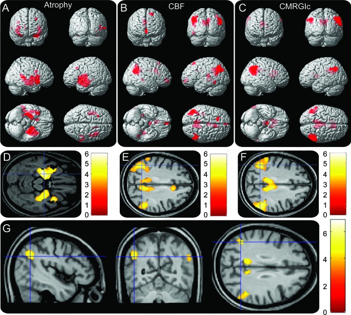
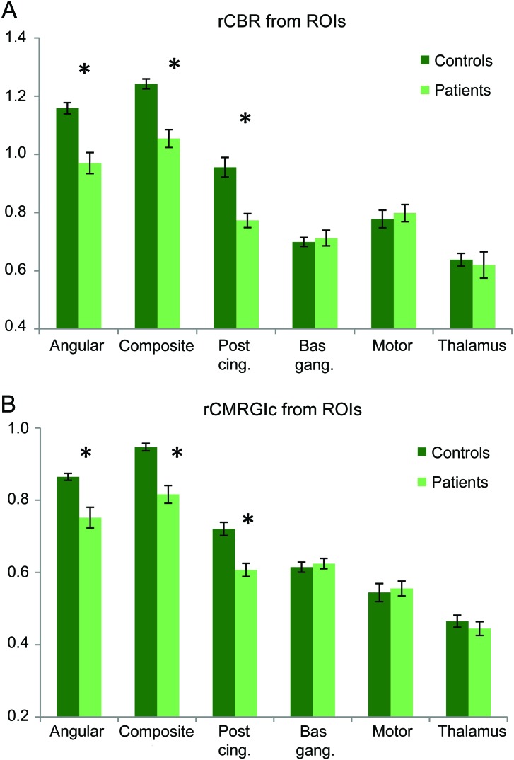
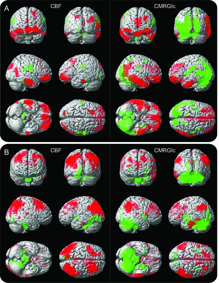
Results: Good agreement between hypoperfusion and hypometabolism patterns was observed, with overlap primarily in bilateral angular gyri and posterior cingulate. ROI results showed similar scales of functional deficit between patients and controls in both modalities. Both ASL and FDG-PET were able to distinguish neural networks associated with different neuropsychological tests with good overlap between modalities.
Conclusions: Our voxel-wise results indicated that ASL-MRI provides largely overlapping information with FDG-PET. ROI analysis demonstrated that both modalities detected similar degrees of functional deficits in affected areas. Given its ease of acquisition and noninvasiveness, ASL-MRI may be an appealing alternative for AD studies.
Figures
Comment in
-
Alzheimer disease: Arterial spin-labeled MRI for diagnosis and monitoring of AD.Nat Rev Neurol. 2011 Dec 26;8(1):3. doi: 10.1038/nrneurol.2011.206. Nat Rev Neurol. 2011. PMID: 22198394 No abstract available.
References
-
- Braak H, Braak E. Neuropathological stageing of Alzheimer-related changes. Acta Neuropathol 1991;82:239–259 - PubMed
Publication types
MeSH terms
Substances
Grants and funding
LinkOut - more resources
Full Text Sources
Other Literature Sources
Medical