

"Asymmetric scalloping of the regenerate": a radiological sign of pseudoaneurysm in distraction osteogenesis
- PMID: 22094536
- PMCID: PMC3225571
- DOI: 10.1007/s11751-011-0121-4
"Asymmetric scalloping of the regenerate": a radiological sign of pseudoaneurysm in distraction osteogenesis
Abstract
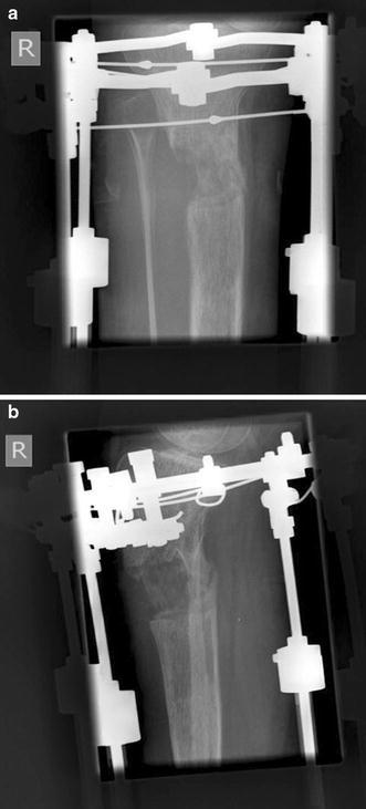
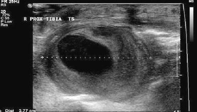
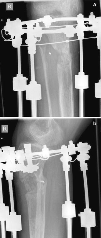
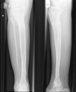
Pseudoaneurysm formation is an uncommon but well-recognised and important complication in limb reconstruction surgery. Postoperative diagnosis is usually clinical or an incidental finding. We present an 11-year-old girl, who underwent two-stage limb lengthening with a circular fixator, for a previously treated pseudoarthrosis of the tibia. During the lengthening plan, a concave defect was noted on one side of the regenerate, which was found to be due to extrinsic compression by a pseudoaneurysm. Normal regenerate formation was seen after selective embolisation of the pseudoaneurysm. This concave appearance on one side of the regenerate has previously been described secondary to a difference in stability on the two sides of the osteotomy, when a monolateral fixator is used, but not due to extrinsic compression by a pseudoaneurysm. The authors propose that this radiographic appearance of "asymmetrical scalloping" on one side of the regenerate may represent a radiological sign of a pseudoaneurysm formation and should provoke investigation for the same.
Figures
References
-
- Maffuli N, Fixsen JA. Distraction osteogenesis in congenital limb length discrepancy: a review. J R Coll Surg Edinb. 1996;41(4):258–264. - PubMed
-
- Endo H, Asaumi K, Mitani S, Noda T, Minagawa H, Tetsunaga T, Ozaki T. The minimally invasive plate osteosynthesis (MIPO) technique with a locking compression plate for femoral lengthening. Acta Med Okayama. 2008;62(5):333–339. - PubMed
LinkOut - more resources
Full Text Sources
