. 2012 Jan 1;72(1):346-55.
doi: 10.1158/0008-5472.CAN-11-2819. Epub 2011 Nov 17.

Mesenchymal stromal cell mutations and wound healing contribute to the etiology of desmoid tumors

Affiliations

Mesenchymal stromal cell mutations and wound healing contribute to the etiology of desmoid tumors

Adelaide M Carothers et al. Cancer Res. .

Abstract

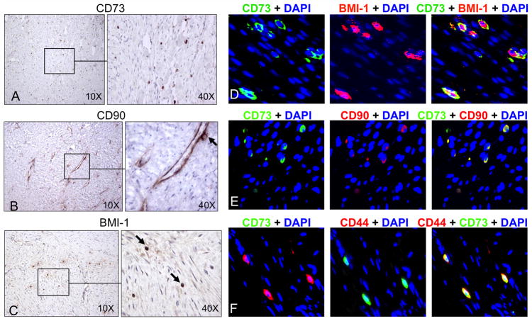
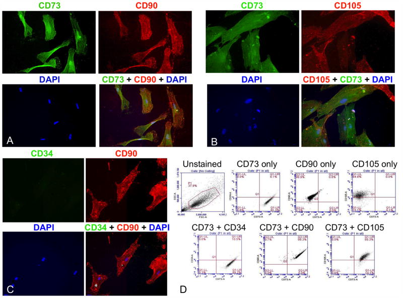
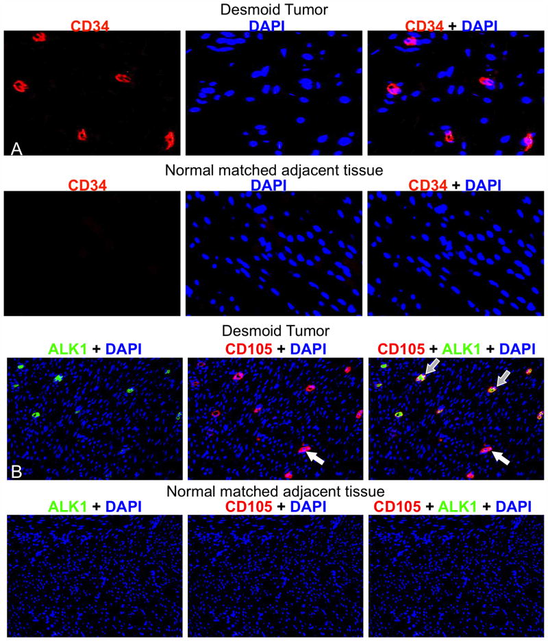
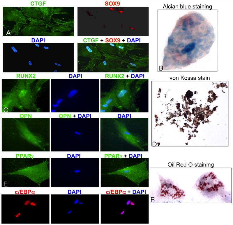
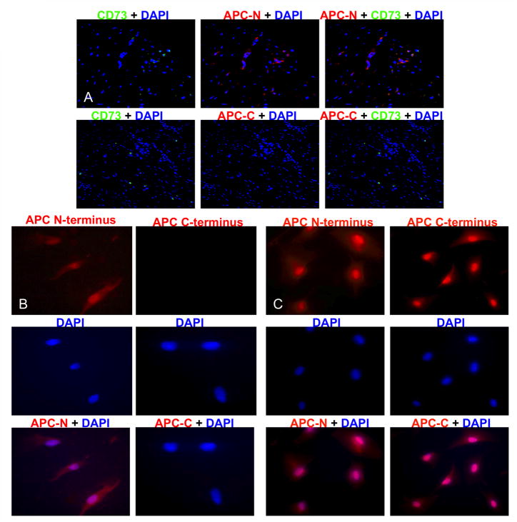
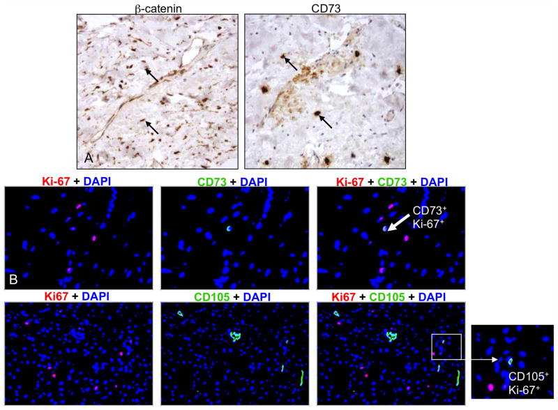
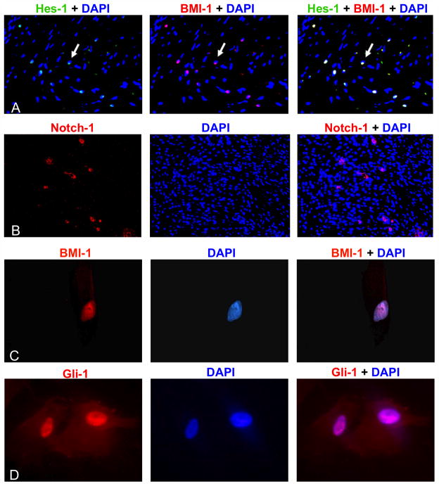
Desmoid tumors are nonmalignant neoplasms of mesenchymal origin that mainly contain fibroblast lineage cells. These tumors often occur in patients with familial adenomatous polyposis (FAP) coli who have germ line mutations in the APC gene. Given emerging data that has implicated multipotent mesencyhmal stromal cells (MSC) in the origin of mesenchymal tumors, we hypothesized that desmoid tumors may arise in patients with FAP after MSCs acquire somatic mutations during the proliferative phase of wound healing. To test this idea, we examined 16 desmoid tumors from FAP-associated and sporadic cases, finding that all 16 of 16 tumors expressed stem cell markers, whereas matching normal stromal tissues were uniformly negative. Desmoid tumors also contained a subclass of fibrocytes linked to wound healing, angiogenesis, and fibrosis. Using an MSC cell line derived from an FAP-associated desmoid tumor, we confirmed an expected loss in the expression of adenomatous polyposis coli (APC) and the transcriptional repressor BMI-1 while documenting the coexpression of markers for chondrocytes, adipocytes, and osteocytes. Together, our findings argue that desmoid tumors result from the growth of MSCs in a wound healing setting that is associated with deregulated Wnt signaling due to APC loss. The differentiation potential of these MSCs combined with expression of BMI-1, a transcriptional repressor downstream of Hedgehog and Notch signaling, suggests that desmoid tumors may respond to therapies targeting these pathways.


Conflict of interest statement

Disclosure of Potential Conflicts of Interest

The authors indicate no potential conflicts of interest.

Figures

Figure 1
Figure 1. Immunohistochemical staining demonstrated stem cell marker expression in DTs
Sixteen archived DTs were stained for stem cell markers CD73 (A), CD90 (B), and BMI-1 (C), and representative images demonstrated the presence of abundant MSCs (black arrows) within tumors (original magnification 10X or 40X). Fluorescent immunohistochemistry of paraffin-embedded DTs showed co-expression of the MSC markers CD73 (green) and BMI-1 (red) (D), CD73 (green) and CD90 (red) (E), as well as CD73 (green) and CD44 (red) (F). Co-expression of both markers produced a yellow overlay. Original magnification was 40X. In this and subsequent fluorescent immunohistochemistry, nuclei were counterstained blue with DAPI.
Figure 2
Figure 2. DT-derived MSCs co-expressed positive but not negative MSC markers
Fluorescent immunohistochemistry was performed on early passage DT-derived MSCs demonstrating co-expression of the MSC markers CD73 (green) and CD90 (red) (A) as well as CD73 (green) and CD105 (red) (B), but not expression of the negative endothelial precursor marker, CD34 (C). Original magnification was 40X. FACS analysis of these cells showed co-expression of the obligate MSC markers CD73, CD90, and CD105, but not CD34 (D). The gated population is outlined in red on the top, left dot plot. Sorting showed that: 99.1% of cells were CD73+, CD34; 99.3% were CD73+, CD90+; and 89.3% were CD73+, CD105+.
Figure 3
Figure 3. DTs contained CD34+, CD105+, and ALK1+ cells
Representative fluorescent immunohistochemistry images are shown of formalin-fixed, paraffin-embedded DTs stained for CD34 (red) (A, Top). Matched normal adjacent tissue did not display any CD34+ cells (A, Bottom) (original magnification 40X). Representative fluorescent immunohistochemistry images of formalin-fixed paraffin-embedded archival DTs stained for ALK1 (red) and CD105 (green) (B, Top). Matched normal adjacent tissue showed only AKL1 and CD105 cells (B, Bottom). Open arrows indicate CD105+ ALK1 cells; grey filled arrows indicate dual positive cells. The original magnification was 20X.
Figure 4
Figure 4. DT-derived MSCs exhibited tri-potent differentiation capability
DT-derived cells were incubated in differentiation media, and confirmation of differentiation was obtained by expression of chondrocyte markers CTGF (green) and Sox9 (red), overlay shows yellow stain (A) plus positive Alcian Blue staining (B); osteocyte markers RUNX2 (green) and OPN (green) (C) plus positive von Kossa staining (D); and adipocyte markers PPARγ (green) and c/EBPα (red) (E) plus positive Oil Red O staining (F). Original magnification was 40X.
Figure 5
Figure 5. MSCs expressed only the APC-N terminus consistent with somatic APC+ LOH
Fluorescent immunohistochemistry was performed on paraffin-embedded DTs and DT-derived cells for both the APC (N-15) and (C-20) termini. Normal human adipose-derived MSCs were used as controls. In MSCs from both the DT (A) and cell line (B), staining was positive for the N-terminus (red) but not the C-terminus. In contrast, staining was positive for both N- and C-termini of APC protein in normal human adipose-derived MSCs (C). Original magnification was 10X and 20X. Overlay shows a purple nuclear stain.
Figure 6
Figure 6. MSCs demonstrated nuclear β-catenin localization
Immunohistochemistry was performed on paraffin-embedded DTs and showed accumulation of nuclear β-catenin in MSCs, indicating active Wnt signaling (A). Original magnification was 10X and 40X. Fluorescent immunohistochemistry of DT sections stained for Ki-67 (red) and CD73 or CD105 (green) identified dually positive cells (B).
Figure 7
Figure 7. Notch and Hedgehog pathways were upregulated in DTs
Fluorescent immunohistochemistry was performed on paraffin-embedded DTs and showed positive expression of the Notch target gene Hes-1 (green) (A) and Notch-1 (red) (B). Co-localization with BMI-1 (red) produced yellow nuclei (A). DT-derived cells also expressed the transcriptional repressor BMI-1 (C). Co-localization of red staining of BMI-1 with the blue counter-stain produced purple-colored nuclei. Similarly, purple nuclei were evident when the MSCs were stained with antibodies for the Hedgehog transcriptional activator, Gli-1 (red) (D).

References

    1. Klemmer S, Pascoe L, DeCosse J. Occurrence of desmoids in patients with familial adenomatous polyposis of the colon. Am J Med Genet. 1987;28:385–392. - PubMed
    1. Rodriquez-Bigas MA, Mahoney MC, Karakousis CP, Petrelli NJ. Desmoid tumors in patients with familial adenomatous polyposis. Cancer. 1994;74:1270–1274. - PubMed
    1. Hansmann A, Adolph C, Vogel T, Unger A, Moeslein G. High-dose tamoxifen and sulindac as first-line treatment for desmoid tumors. Cancer. 2004;100:612–620. - PubMed
    1. Mace J, Sybil Biermann J, Sondak V, McGinn C, Hayes C, Thomas D, et al. Response of extra-abdominal desmoid tumors to therapy with imatinib mesylate. Cancer. 2002;95:2373–2379. - PubMed
    1. Bertagnolli MM, Morgan JA, Fletcher CD, Raut CP, Dileo P, Gill RR, et al. Multimodality treatment of mesenteric desmoid tumors. Eur J Cancer. 2008;44:2404–2410. - PubMed

Publication types