. 2012 Feb;190(2):511-21.
doi: 10.1534/genetics.111.134080. Epub 2011 Nov 17.

A conserved transcriptional regulator governs fungal morphology in widely diverged species

Affiliations

A conserved transcriptional regulator governs fungal morphology in widely diverged species

Christopher W Cain et al. Genetics. 2012 Feb.

Abstract

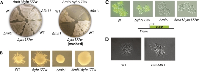
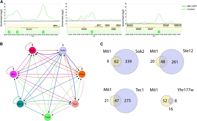
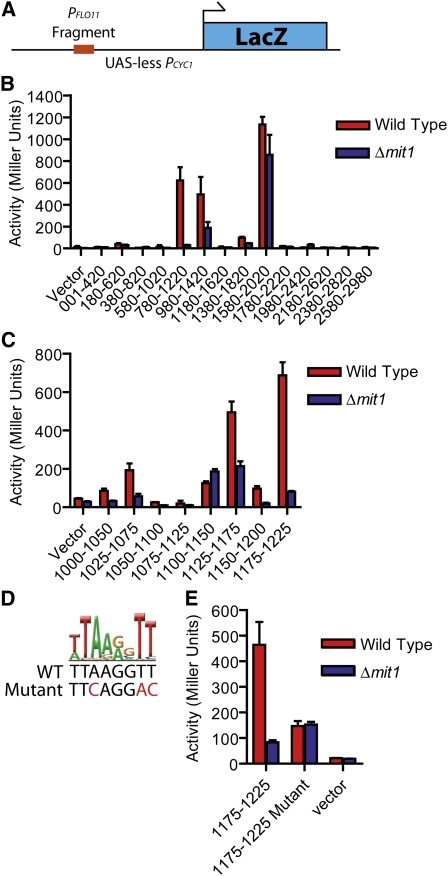
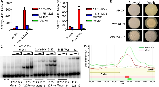
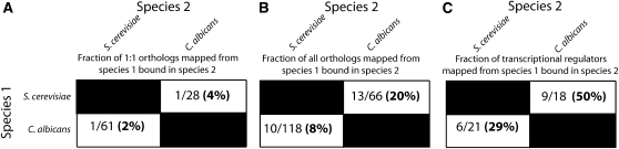
Fungi exhibit a large variety of morphological forms. Here, we examine the functions of a deeply conserved regulator of morphology in three fungal species: Saccharomyces cerevisiae, Candida albicans, and Histoplasma capsulatum. We show that, despite an estimated 600 million years since those species diverged from a common ancestor, Wor1 in C. albicans, Ryp1 in H. capsulatum, and Mit1 in S. cerevisiae are transcriptional regulators that recognize the same DNA sequence. Previous work established that Wor1 regulates white-opaque switching in C. albicans and that its ortholog Ryp1 regulates the yeast to mycelial transition in H. capsulatum. Here we show that the ortholog Mit1 in S. cerevisiae is also a master regulator of a morphological transition, in this case pseudohyphal growth. Full-genome chromatin immunoprecipitation experiments show that Mit1 binds to the control regions of the previously known regulators of pseudohyphal growth as well as those of many additional genes. Through a comparison of binding sites for Mit1 in S. cerevisiae, Wor1 in C. albicans, and Wor1 ectopically expressed in S. cerevisiae, we conclude that the genes controlled by the orthologous regulators overlap only slightly between these two species despite the fact that the DNA binding specificity of the regulators has remained largely unchanged. We suggest that the ancestral Wor1/Mit1/Ryp1 protein controlled aspects of cell morphology and that movement of genes in and out of the Wor1/Mit1/Ryp1 regulon is responsible, in part, for the differences of morphological forms among these species.


Figures

Figure 1
Figure 1
MIT1 is required for haploid invasive growth, diploid pseudohyphal growth, and expression of FLO11 in S. cerevisiae. (A) Haploid invasive growth assay. Haploid a cells of the indicated genotypes (Σ2000 background) were plated on YPD agar plates, grown for 2 days at 30°, and washed. Pre- (left) and post- (right) wash images are shown. (B) Diploid pseudohyphal growth assay. Individual a/α diploid cells (Σ2000 background) were plated on SLAD agar plates, grown for 7 days, and then photographed. A representative colony from each plate is shown. Δmit1 and Δmit1Δyhr177w colonies were never observed to display pseudohyphal growth in this assay. (C) Expression of FLO11 is dependent on MIT1. The FLO11 coding sequence was replaced with GFP (S65T). Haploid a cells were grown in YPD to log phase, placed under a coverslip, and then photographed at 40× magnification. (D) MIT1 expressed from the TEF promoter drives diploid pseudohyphal growth in SD media conditions. Individual diploid Σ2000 cells were plated on an SD agar pad on a glass microscopy slide and allowed to grow under a coverslip at 30° for 16 hr, and then individual microcolonies were visualized at 40× magnification. Wild-type cells were not observed to display pseudohyphal growth while cells containing a plasmid expressing MIT1 from the TEF promoter displayed elongated morphology and agar penetration consistent with the pseudohyphal growth phenotype.
Figure 2
Figure 2
MIT1 is a central transcriptional regulator of filamentous growth. (A) ChIP-chip plots illustrate binding of Mit1-GFP in Σ2000 haploid a cells to promoters of FLO11, MIT1, and SOK2. Experimental enrichment data are presented as a solid green line in each panel and control data from a wild-type untagged strain are presented as a dashed red line. The green boxes in the bottom track represent the called Mit1 binding sites. Data were visualized using Mochiview 1.45, with the x-axis representing the genomic location and the y-axis representing log2 enrichment. (B) The network of interactions between Mit1 and previously known diploid pseudohyphal growth regulators. Arrows indicate binding of a regulator at the promoter of a given regulator. Connections represent those previously reported for the other six regulators (Borneman et al. 2006, 2007b) and those from the Mit1 ChIP-chip data reported here. (C) A genome-wide binding analysis comparison of Mit1 and Yhr177w bound intergenic regions with those previously reported for Sok2, Ste12, and Tec1 (Borneman et al. 2007b) reveals a high degree of target gene overlap.
Figure 3
Figure 3
Mit1 acts on a discrete location within the FLO11 promoter corresponding to the Mit1/Wor1 DNA-binding motif. (A) Schematic of the UAS-less PCYC1 plasmid with a FLO11 promoter fragment used for the β-galactosidase activation assays. (B) Fragments (440 bp) of the FLO11 promoter were inserted into a UAS-less CYC1 promoter upstream of a LacZ reporter construct, as previously reported (Rupp et al. 1999). These plasmids were transformed into either wild-type haploid a cells (red) or Δmit1 haploid a cells (blue), and standard β-galactosidase assays were performed in selective SD media. β-Galactosidase units of activity (Miller units) are plotted on the y-axis, and fragments of the FLO11 promoter are indicated on the x-axis (numbers represent the position of individual fragments relative to the FLO11 start codon). (C) Fragments (50 bp) of the FLO11 promoter were inserted into the LacZ reporter plasmid and β-galactosidase activity was measured for strains grown under the same conditions as in B. Fragment 1175–1225 displayed the strongest activity, and this activity was Mit1 dependent. Axis labeling is as in B. (D and E) A sequence resembling the Mit1/Wor1 recognition motif (displayed as a logo) was identified in the 1175–1225 FLO11 promoter fragment. Three bases of this motif were mutated as shown (red), and the corresponding plasmid (“1175–1225 Mutant”) exhibited reduced Mit1-dependent β-galactosidase activity when transformed into yeast. Axis labeling is as in B. Error bars in B, C, and E represent the standard deviation from two or three experiments.
Figure 4
Figure 4
DNA binding and activation functions are conserved among S. cerevisiae Mit1, C. albicans Wor1, and H. capsulatum Ryp1. (A and B) Reporter plasmids carrying a functional upstream 1175–1225 FLO11 promoter fragment (1175–1225, red), the nonfunctional mutated version of the fragment (1175–1225 mutant, blue), or no insert (vector, green) were cotransformed with a plasmid containing the TEF promoter driving an empty vector, (A) C. albicans WOR1, or (B) H. capsulatum RYP1. Both PTEF-WOR1 and PTEF-RYP1 activate transcription from the wild-type but not the mutant FLO11 fragment-CYC1 construct. β-Galactosidase units of activity (Miller units) are plotted on the y-axis, and error bars in A and B represent the standard deviation from two or three experiments. (C) Yhr177w, Mit1, and Wor1 bind to the 1175–1225 fragment from the FLO11 promoter, as monitored by gel shift assays. Mit1 (5–251 aa), Yhr177w (6–201 aa), and MBP-Wor1 (1–321 aa) were incubated with either the FLO11 1175–1225 promoter fragment (1175–1225, +) or the FLO11 1175–1225 mutant fragment (1175–1225 mutant, −). All three proteins bind to the FLO11 1175–1225 promoter fragment but not to the mutated fragment, indicating that the binding of Mit1, Yhr177w, and Wor1 to the FLO11 promoter is sequence specific. Yhr177w and Mit1 concentrations range from 0.5 to 4 nM, and MBP-Wor1 concentrations range from 2 to 8 nM. (D) A ChIP-chip plot showing ectopically expressed Wor1 associating with the FLO11 promoter in vivo. Wor1 was immunoprecipitated from a Δmit1Δyhr177w strain carrying the PTEF-WOR1 plasmid (solid red line), and the control is a Δmit1Δyhr177w strain carrying PTEF-vector plasmid (dashed red line). ChIP data for the Mit1-GFP experiments in a Mit1-GFP strain (solid green line) or in a control strain lacking GFP (dashed green line) are included for comparison. The red box in the bottom track represents a called Wor1 binding site and the green box represents a called Mit1 binding site. Data were visualized using Mochiview 1.45, with the x-axis representing the genomic location and the y-axis representing log2 enrichment. (E) Ectopic expression of WOR1 or RYP1 induces invasive growth on SD media. a/α diploid Σ2000 cells were spotted on SD agar, grown for 2 days at 30°, and then washed under a stream of water. Pre- (left) and post- (right) wash images are shown.
Figure 5
Figure 5
A comparison of the Mit1 regulon in S. cerevisiae to the Wor1 regulon in C. albicans indicates there is a small conserved set of orthologous genes that code for transcriptional regulators that are bound by both Mit1 and Wor1. (A–C) The number of Mit1- or Wor1-bound genes in species 1 that are also bound in species 2, as a fraction of the total genes bound in species 1 that can be mapped to orthologs in species 2 when (A) only 1:1 orthologous relationships are considered, (B) all orthologous relationships (i.e., 1:1, 2:1, 3:5) are considered, or (C) only Mit1- or Wor1-bound genes that encode sequence-specific DNA-binding proteins are considered.

References

    1. Anderson J. M., Soll D. R., 1987. Unique phenotype of opaque cells in the white-opaque transition of Candida albicans. J. Bacteriol. 169: 5579–5588 - PMC - PubMed
    1. Baker C. R., Tuch B. B., Johnson A. D., 2011. Extensive DNA-binding specificity divergence of a conserved transcription regulator. Proc. Natl. Acad. Sci. USA 108: 7493–7498 - PMC - PubMed
    1. Borneman A. R., Leigh-Bell J. A., Yu H., Bertone P., Gerstein M., et al. , 2006. Target hub proteins serve as master regulators of development in yeast. Genes Dev. 20: 435–438 - PMC - PubMed
    1. Borneman A. R., Gianoulis T. A., Zhang Z. D., Yu H., Rozowsky J., et al. , 2007a Divergence of transcription factor binding sites across related yeast species. Science 317: 815–819 - PubMed
    1. Borneman A. R., Zhang Z. D., Rozowsky J., Seringhaus M. R., Gerstein M., et al. , 2007b Transcription factor binding site identification in yeast: a comparison of high-density oligonucleotide and PCR-based microarray platforms. Funct. Integr. Genomics 7: 335–345 - PubMed

Publication types

MeSH terms