. 2012 Feb;8(Suppl 1):3-26.
doi: 10.1007/s11302-011-9271-6. Epub 2011 Nov 18.

In pursuit of P2X3 antagonists: novel therapeutics for chronic pain and afferent sensitization

Affiliations

In pursuit of P2X3 antagonists: novel therapeutics for chronic pain and afferent sensitization

Anthony P Ford. Purinergic Signal. 2012 Feb.

Abstract

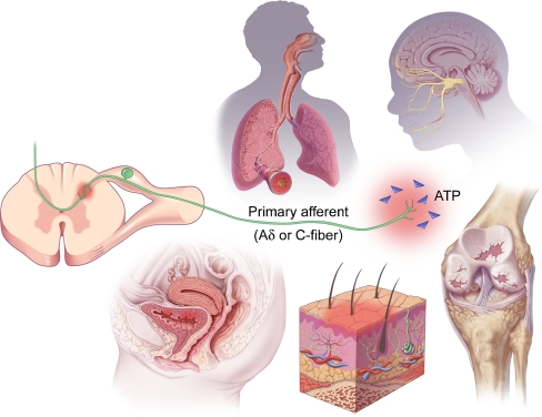
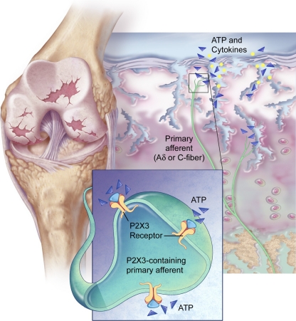
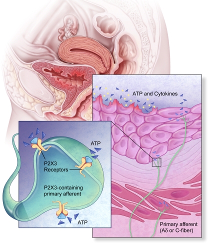
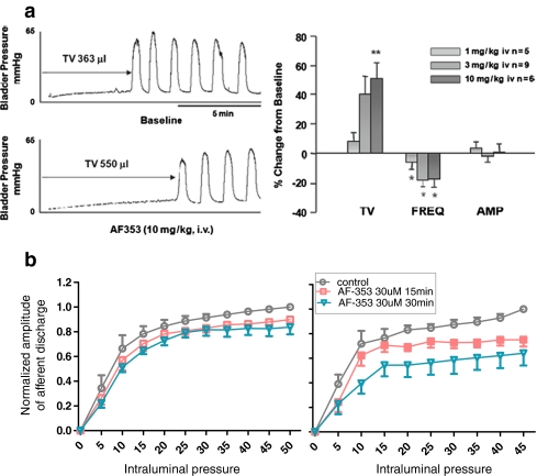
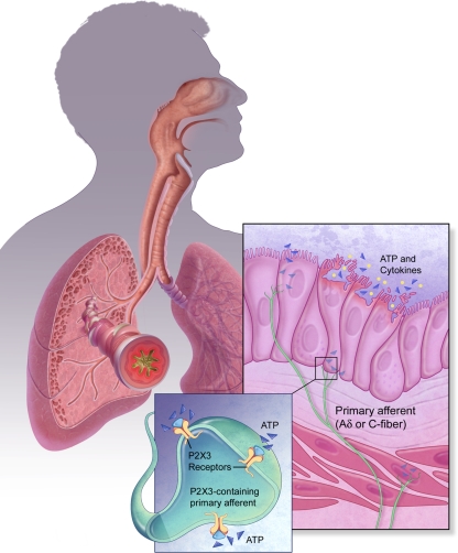
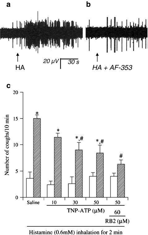
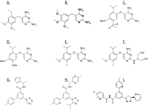
Treating pain by inhibiting ATP activation of P2X3-containing receptors heralds an exciting new approach to pain management, and Afferent's program marks the vanguard in a new class of drugs poised to explore this approach to meet the significant unmet needs in pain management. P2X3 receptor subunits are expressed predominately and selectively in so-called C- and Aδ-fiber primary afferent neurons in most tissues and organ systems, including skin, joints, and hollow organs, suggesting a high degree of specificity to the pain sensing system in the human body. P2X3 antagonists block the activation of these fibers by ATP and stand to offer an alternative approach to the management of pain and discomfort. In addition, P2X3 is expressed pre-synaptically at central terminals of C-fiber afferent neurons, where ATP further sensitizes transmission of painful signals. As a result of the selectivity of the expression of P2X3, there is a lower likelihood of adverse effects in the brain, gastrointestinal, or cardiovascular tissues, effects which remain limiting factors for many existing pain therapeutics. In the periphery, ATP (the factor that triggers P2X3 receptor activation) can be released from various cells as a result of tissue inflammation, injury or stress, as well as visceral organ distension, and stimulate these local nociceptors. The P2X3 receptor rationale has aroused a formidable level of investigation producing many reports that clarify the potential role of ATP as a pain mediator, in chronic sensitized states in particular, and has piqued the interest of pharmaceutical companies. P2X receptor-mediated afferent activation has been implicated in inflammatory, visceral, and neuropathic pain states, as well as in airways hyperreactivity, migraine, itch, and cancer pain. It is well appreciated that oftentimes new mechanisms translate poorly from models into clinical efficacy and effectiveness; however, the breadth of activity seen from P2X3 inhibition in models offers a realistic chance that this novel mechanism to inhibit afferent nerve sensitization may find its place in the sun and bring some merciful relief to the torment of persistent discomfort and pain. The development philosophy at Afferent is to conduct proof of concept patient studies and best identify target patient groups that may benefit from this new intervention.


Figures

Fig. 1
Fig. 1
P2X3 containing ionotropic receptors are found in a large proportion of unmyelinated and thinly myelinated primary afferent nerves innervating essentially all tissues and organs. ATP is released from many cell types in these receptive fields, as well as at the central terminals of activated afferents, and more so under conditions of injury, inflammation, stress, movement, and distension. Afferent sensitization results and appears to contribute to the serious symptoms of acute and chronic pain and irritation in musculoskeletal, visceral and neuropathic sensory disorders
Fig. 2
Fig. 2
Pain from damaged and strained joints and other musculoskeletal structures chronically impacts the lives of a vast number of individuals. Available pain medicines are often inadequately effective, poorly tolerated or considered unsafe, highlighting the great demand for medicines with novel mechanisms of action. P2X3 antagonists have shown to be effective in reversing hyperalgesia in several animal models of joint pain, and in studies of pain from cancer invasion of bony tissues. Osteoarthritis of the knee, an exceptionally common condition, is one of several high potential indications for novel P2X3 antagonists
Fig. 3
Fig. 3
Nocifensive data showing the effect of a P2X3 antagonist in preclinical models of joint hyperalgesia. a In an adjuvant-induced arthritis model in rat (7d following intraplantar administration of complete Freund's adjuvant), AF-353 produces dose-dependent anti-hyperalgesia in weight-bearing asymmetry and von Frey filament mechanical tests; magnitude of effect is compared with that of the NSAID naproxen. b In a rat model of knee osteoarthritis (14d following intra-articular administration of monoiodoacetate), AF-353 produces dose-dependent anti-hyperalgesia in weight-bearing asymmetry test (left; compared with the COX-2 inhibitor Vioxx). In the same model, following repeated dosing, AF-219 (7d bid, orally; right) attenuates the weight bearing laterality with complete reversal of apparent hyperalgesia at the two higher doses
Fig. 4
Fig. 4
Systemic P2X3 receptor antagonism with AF-353 attenuates bone cancer pain behaviour in rats. Intra-tibial injection of MRMT-1 carcinoma cells (black circle) induced significant increases in both mechanical allodynia (a and c) and weight-bearing difference (b and d) compared with the control rats (yellow square and open triangle). Oral administration of AF-353 (bid) significantly attenuated both MRMT-1 carcinoma cell-induced mechanical allodynia and weight-bearing difference when given before development of bone cancer pain behavior (termed prophylactic treatment, red circle), while bone cancer-induced established mechanical allodynia could be significantly reduced by AF-353 (termed reversal treatment: diamond). The red and blue bars represent the prophylactic and reversal treatment dosing periods, respectively. Mechanical allodynia was quantified by calculating the area under the curve values determined from a plot of the percentage of positive ipsilateral withdrawal response at each filament against the filament force on a logarithmic scale. Hindlimb weight-bearing difference was expressed as contralateral minus ipsilateral readings. Values are expressed as means ± SEM. **P < 0.01 and ***P < 0.001 versus the cancer + vehicle group (from [66]). In the lower panel (e) is shown immunohistochemical characterization of the retrogradely labeled Fast Blue DRG neurons innervating the rat tibia. Fast Blue (blue) labeling was present in both P2X3- (green) and calcitonin gene-related peptide (CGRP)-positive (red) neurons at the L2 and L3 DRG levels
Fig. 5
Fig. 5
Sensory fibers in visceral organs, especially the urinary bladder, express high levels of P2X3 receptors that are elevated in pathological conditions; unmasking segmental spinal reflexes that sense ATP content during filling and distension. P2X3 antagonists suppress afferent excitation and raise filling volume thresholds, especially in rodent models of cystitis. The distressing and largely unmet painful and irritative symptoms of bladder pain syndrome/interstitial cystitis and chronic prostatitis as well as lower urinary tract symptoms (urgency, frequency, and nocturia) associated with overactive bladder and benign prostatic hyperplasia, represent important visceral indications for novel P2X3 antagonists
Fig. 6
Fig. 6
Effects of P2X3 antagonists in preclinical models of urinary bladder reflexes. a P2X3 antagonism (AF-353) significantly raises volume thresholds and lowers frequency of micturition reflexes in filling cystometry models in rats anesthetized with urethane, which unmasks segmental spinal pathways. b Bladder–pelvic nerve preparation from cyclophosphamide (CPM) sensitized rats. Afferent nerve firing associated with volume expansion is markedly reduced by P2X3 antagonism with AF-353, applied either to serosal (extraluminal, left) or intravesical (intraluminal right) surface of the bladder (figures kindly provided by Prof Weifang Rong, Shanghai Jiaotong University, China)
Fig. 7
Fig. 7
Airway sensitization and irritation. Large proportions of C and Aδ fiber afferents in the upper and lower airways express P2X3 receptors and can be sensitized by ATP. Activation of some of these sensory nerves by ATP liberated from epithelial and smooth muscle cells is postulated to contribute to components of airways hyperexcitability, giving rise to airways hypersecretion, bronchospasm, breathlessness (dyspnea), and chronic cough, all of which are undermanaged factors in chronic diseases such as COPD and asthma
Fig. 8
Fig. 8
a, b Extracellular recording of action potential discharge from an intrapulmonary nodose C-fiber in an ex vivo isolated, perfused lung-nerve preparation from guinea pig. a Representative recording of action potential discharge from a single lung nodose C-fiber (conduction velocity 0.78 m/s) in response to histamine (HA, 30 μM) delivered as a 1 ml bolus injection into both the pulmonary artery and the trachea. b Perfusion of the same C-fiber with AF-353 (100 μM, 15 min) reduced both the peak frequency of action potential discharge and the total number of action potentials (AP) generated in response to HA by 64% (figures kindly provided by Prof Brad Undem, Johns Hopkins University, USA). c Effect of TNP-ATP on the histamine-induced increase in the number of citric acid-induced coughs in guinea pigs. The guinea pigs were exposed to 0.6 mM histamine aerosol for 2 min during the 5 min preceding the inhalation of 0.1 M citric acid. Guinea pigs were exposed to TNP-ATP or reactive blue 2 (RB2) for 2 min during the 5 min preceding the inhalation of histamine. The number of coughs during 10 min of exposure to citric acid was counted before (open column) and after exposure to histamine (hatched column). Each column represents the mean with S.E.M. for five animals. *P < 0.05 vs. the value before exposure to histamine. #P < 0.05 vs. the value after exposure to histamine of saline-treated guinea pigs (reproduced with permission from [117])
Fig. 9
Fig. 9
Chemical structures of various key compounds that antagonize P2X3 containing receptors: a AF-001 (original DAP HTS “hit”, P2X3 IC50 1.4 μM); b trimethoprim (inactive at 30 μM); c AF-130 (RO-13; 100 nM); d AF-010 (RO-10; 40 nM); e AF-353 (RO-4; 6 nM); f AF-906 (RO-51; 2 nM); g AF-454 (original SAA HTS “hit”, 0.65 μM) ; H, AF-014 (3 nM); I, MK-3901 (Merck lead published at ACS 2011; 24 nM)

References

    1. Burnstock G, Fredholm BB, North RA, Verkhratsky A. The birth and postnatal development of purinergic signalling. Acta Physiol. 2010;199(2):93–147. doi: 10.1111/j.1748-1716.2010.02114.x. - DOI - PubMed
    1. Gunosewoyo H, Kassiou M. P2X purinergic receptor ligands: recently patented compounds. Expert Opin Ther Pat. 2010;20:625–646. doi: 10.1517/13543771003702424. - DOI - PubMed
    1. Gunosewoyo H, Coster MJ, Kassiou M. Molecular probes for P2X7 receptor studies. Curr Med Chem. 2007;14(14):1505–1523. doi: 10.2174/092986707780831023. - DOI - PubMed
    1. Romagnoli R, Baraldi PG, Cruz-Lopez O, et al. The P2X7 receptor as a therapeutic target. Expert Opin Ther Targets. 2008;12(5):647–661. doi: 10.1517/14728222.12.5.647. - DOI - PubMed
    1. Carroll WA, Donnelly-Roberts DL, Jarvis MF. Selective P2X7 receptor antagonists for chronic inflammation and pain. Purinergic Signal. 2009;5:63–73. doi: 10.1007/s11302-008-9110-6. - DOI - PMC - PubMed