. 2012 Mar 1;185(5):537-46.
doi: 10.1164/rccm.201106-0965OC. Epub 2011 Nov 17.

Regulation of transforming growth factor-β1-driven lung fibrosis by galectin-3

Affiliations

Regulation of transforming growth factor-β1-driven lung fibrosis by galectin-3

Alison C Mackinnon et al. Am J Respir Crit Care Med. .

Abstract

Rationale: Idiopathic pulmonary fibrosis (IPF) is a chronic dysregulated response to alveolar epithelial injury with differentiation of epithelial cells and fibroblasts into matrix-secreting myofibroblasts resulting in lung scaring. The prognosis is poor and there are no effective therapies or reliable biomarkers. Galectin-3 is a β-galactoside binding lectin that is highly expressed in fibrotic tissue of diverse etiologies.

Objectives: To examine the role of galectin-3 in pulmonary fibrosis.

Methods: We used genetic deletion and pharmacologic inhibition in well-characterized murine models of lung fibrosis. Further mechanistic studies were performed in vitro and on samples from patients with IPF.

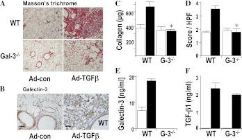
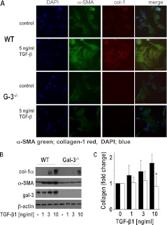
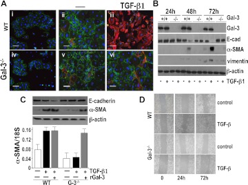
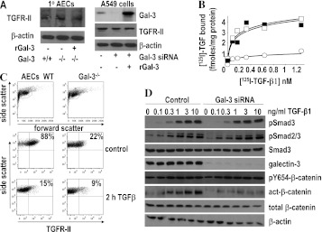
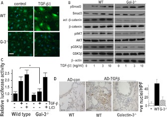
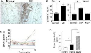
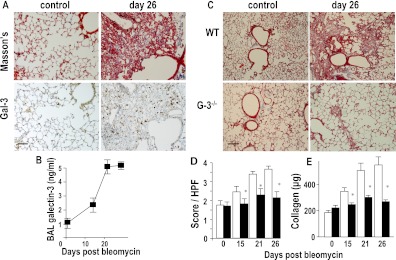
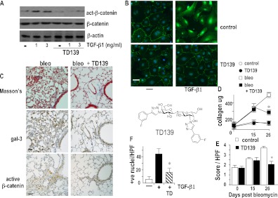
Measurements and main results: Transforming growth factor (TGF)-β and bleomycin-induced lung fibrosis was dramatically reduced in mice deficient in galectin-3, manifest by reduced TGF-β1-induced EMT and myofibroblast activation and collagen production. Galectin-3 reduced phosphorylation and nuclear translocation of β-catenin but had no effect on Smad2/3 phosphorylation. A novel inhibitor of galectin-3, TD139, blocked TGF-β-induced β-catenin activation in vitro and in vivo and attenuated the late-stage progression of lung fibrosis after bleomycin. There was increased expression of galectin-3 in the bronchoalveolar lavage fluid and serum from patients with stable IPF compared with nonspecific interstitial pneumonitis and controls, which rose sharply during an acute exacerbation suggesting that galectin-3 may be a marker of active fibrosis in IPF and that strategies that block galectin-3 may be effective in treating acute fibrotic exacerbations of IPF.

Conclusions: This study identifies galectin-3 as an important regulator of lung fibrosis and provides a proof of principle for galectin-3 inhibition as a potential novel therapeutic strategy for IPF.


Figures

Figure 1.
Figure 1.
Galectin-3−/− mice show reduced lung fibrosis after intratracheal adenoviral transforming growth factor (TGF)-β1 compared with wild-type (WT) mice. WT C57/Bl6 mice or galectin-3−/− mice (G-3−/−) received 2 × 10⁸ plaque-forming units (PFU) adenoviral TGF-β1 (Ad-TGF-β) or control virus (control) and lungs were harvested after 14 days. (A) Lung sections were stained with Masson trichrome (scale bar = 100 μm). Pictures are representative of n = 6 per group. (B) Serial lung sections were immunostained for galectin-3 and showed spatial localization within fibrotic areas of lung (scale bar = 20 μm). (C) Total lung collagen was quantified by sircol assay. (D) Fibrosis was quantified by histologic score from Masson-stained sections. (E) Galectin-3 was increased in the bronchoalveolar lavage fluid from WT mice after Ad-TGF-β. (F) TGF-β1 levels were increased in the bronchoalveolar lavage fluid from WT and galectin-3−/− mice after Ad-TGF-β. n = 6 per group. * P < 0.01 compared with WT.
Figure 2.
Figure 2.
Galectin-3−/− primary lung fibroblasts show reduced collagen production in response to transforming growth factor (TGF)-β1 compared with wild-type (WT) fibroblasts. Primary lung fibroblasts from WT and galectin-3−/− lungs were incubated with or without 5 ng/ml TGF-β1 for 48 hours. (A) Immunofluorescence staining for α-smooth muscle actin (α-SMA) (green) and collagen-1 (red) showing reduced α-SMA and collagen-1 expression in galectin-3−/− fibroblasts after TGF-β1 (scale bar = 15 μm). Nuclei are stained with 4′,6-diamidino-2-phenylindole (DAPI, blue). (B) Reduced a-SMA and collagen-1 expression in TGF-β1–treated galectin-3−/− fibroblasts compared with WT fibroblasts as measured by Western blot analysis. (C) Galectin-3−/− lung fibroblasts produced less collagen compared with WT fibroblasts as determined by sircol assay (n = 4; * P < 0.05 compared with WT).
Figure 3.
Figure 3.
Primary lung alveolar epithelial cells (AEC) from galectin-3−/− mice show reduced epithelial to mesenchymal transition (EMT) in response to transforming growth factor (TGF)-β1 in vitro compared with wild-type (WT) AECs. (A) WT and galectin-3−/− (G-3−/−) AECs 3 days after isolation were untreated (Day 0; i and iv) or cultured for 48 hours in the presence (iii and vi) or absence (ii and v) of 5 ng/ml TGF-β1. Cells were examined by immunofluorescence for E-cadherin (red) and α-smooth muscle actin (α-SMA) (green) expression (scale bar = 100 μm). (B) Primary AECs treated with TGF-β1 were lysed and analyzed for galectin-3, α-SMA, vimentin, E-cadherin, and β-actin expression by Western blot analysis. (C) Galectin-3−/− AECs were cultured for 48 hours with 5 ng/ml TGF-β1 in the presence or absence of 25 μg/ml recombinant galectin-3 and compared with WT AECs treated with TGF-β1. α-SMA, E-cadherin, and β-actin protein expression was analyzed by Western blot analysis and α-SMA transcript expression was analyzed by reverse transcriptase polymerase chain reaction. (n = 4). (D) Confluent cultures of WT and galectin-3−/− lung epithelial cells were wounded and treated with or without 5 ng/ml TGF-β1. Migration into the scored region was observed by phase contrast microscopy.
Figure 4.
Figure 4.
The effect of galectin-3 on transforming growth factor (TGF)-β receptor binding, Smad, and β-catenin signaling in A549 alveolar epithelial cells (AEC). (A) Western blot analysis of total TGFR-II in whole cell lysates from wild-type and galectin-3−/− primary AEC and human A549 cells treated with and without galectin-3 siRNA. (B) Galectin-3 expression was inhibited by siRNA in human A549 cells and cells were treated with 125I-TGF-β1 for 3 hours at 4°C, washed, and binding determined by scintillation counting (solid squares). Inhibition of galectin-3 expression reduced 125I–TGF-β receptor binding by a reduction in Bmax with no significant effect on TGF-β1 binding affinity (open circles). Receptor binding was restored when cells were pretreated for 18 hours with 25 μg/ml recombinant galectin-3 (open squares). (C) TGFR-II expression measured by flow cytometry in control and galectin-3–depleted cells treated with or without 10 ng/ml TGF-β1 for 2 hours. (D) Inhibition of galectin-3 expression has no effect on Smad2/3 phosphorylation but reduces β-catenin activation. A549 were transfected with siRNA duplexes targeting galectin-3 and treated with TGF-β1 at concentrations as indicated for 30 minutes. Lysates were immunoblotted for pSmad3, pSmad2/3, total Smad3, Y654-β-catenin, active-β-catenin, total β-catenin, and β-actin. Galectin-3 blots show efficient knockdown of expression in siRNA-treated cells. There was no difference in Smad2 or 3 phosphorylation after galectin-3 inhibition.
Figure 5.
Figure 5.
The effect of galectin-3 on transforming growth factor (TGF)-β1–induced Smad and β-catenin signaling in primary mouse alveolar epithelial cells (AECs). (A) Fluorescence microscopy of primary AECs from wild-type (WT) and galectin-3−/− mouse stimulated with 10 ng/ml TGF-β1 for 48 hours and stained with an anti–β-catenin antibody (green) and DAPI (blue) (scale bar = 10 μm). (B) Primary AECs from WT and galectin-3−/− mouse lungs were stimulated with TGF-β1 for 30 minutes (Smad3) or 48 hours (β-catenin, AKT, GSK-3β) and cell lysates were analyzed for total and phospho-Smad3 and total and active β-catenin, pAKT, and pSer9-GSK-3β by Western blot. (C) WT and galectin-3−/− AECs were transfected with 1 μg TOPflash and FOPflash and treated with 5 ng/ml TGF-β1 or 10 mM LiCl for 24 hours. Results are expressed as relative luciferase activity and represent the mean SEM of four independent experiments. (D) β-Catenin activation in galectin-3−/− mouse lung in vivo. Mice were given adenoviral TGF-β1 intratracheally and lungs harvested after 14 days. Sections from WT and galectin-3−/− mouse lungs mouse were immunostained for active β-catenin (scale bar = 25 μm). Nuclear staining was observed in areas of fibroproliferation in WT mice. Bar chart showing quantification of positively stained cells in five random fields, n = 6 per group. * P < 0.05 compared with WT.
Figure 6.
Figure 6.
Galectin-3 is elevated in the lungs and serum of patients with idiopathic pulmonary fibrosis (IPF). (A) Sections from human usual interstitial pneumonia (UIP) lung and from age-matched control subjects were stained for galectin-3. Galectin-3 is seen within fibroblastic foci in UIP (scale bar = 100 μm). (B) Galectin-3 was measured by ELISA and was elevated in the bronchoalveolar lavage (BAL) fluid (left) and serum (right) from biopsy-proved patients with UIP and nonspecific interstitial pneumonia (NSIP) and compared with age-matched healthy control subjects (n = 10; * P < 0.01 compared with control). (C) Serial serum levels of galectin-3 were measured by ELISA in 16 diagnosed patients with UIP over 12 months. Two patients showed increased serum galectin-3 before an acute exacerbation. (D) Age-matched control subjects (n = 10), patients with stable UIP (n = 10), and patients having an acute exacerbation of UIP (n = 5). * P < 0.01. ** P < 0.001 compared with control.
Figure 7.
Figure 7.
Galectin-3−/− mice show reduced fibrosis after intratracheal bleomycin. (A) Representative sections from mouse lung at Days 0 and 26 days after bleomycin (33 μg intratracheally) stained for collagen (Masson trichrome) and galectin-3. Images are representative of n = 6 mice per group (scale bar = 50 μm). (B) Galectin-3 was measured in bronchoalveolar lavage (BAL) fluid by ELISA at Days 15, 21, and 26 after intratracheal administration of bleomycin. (C) Representative Masson trichrome–stained sections of wild type (WT) and galectin-3−/− mouse lung after bleomycin (scale bar = 100 μm). (D) Semiquantitative fibrosis score from Masson trichrome–stained sections of WT (open bars) and galectin-3−/− (solid bars) mouse lung. (E) Quantification of collagen content of lung homogenate by sircol assay. * P < 0.05 compared with WT.
Figure 8.
Figure 8.
Galectin-3 inhibitor TD139 reduces bleomycin-induced fibrosis. Upper: primary alveolar epithelial cells from wild-type (WT) mice were plated and treated with transforming growth factor (TGF)-β1 in the presence or absence of 10 μM TD139. (A) Cells were lysed and analyzed for active β-catenin, total β-catenin, and β-actin by Western blot. (B) Cells were fixed and analyzed by immunofluorescence staining for active β-catenin (green) nuclei stained with DAPI (scale bar = 15 μm). (C) Mice were given 0.033 mg bleomycin intratracheally and then saline (bleo) or 10 μg TD139 (TD139) was instilled into the lungs on Days 18, 20, 22, and 24 and lungs were harvested on Day 26 as described in Methods. Serial sections of mouse lung were stained with Masson's trichrome, galectin-3, and active β-catenin (scale bar = 20 μm) as indicated. (D) Fibrosis was assessed by lung collagen content by sircol assay (n = 5–6; * P < 0.01 compared with bleomycin alone) and (E) by histologic score. (F) Active β-catenin was scored from sections of WT and TD139-treated WT mouse lungs. Nuclear staining was observed in areas of fibroproliferation in WT mice. Bar chart showing quantification of positively stained cells in five random fields, n = 6 per group. Results are representative of at least four individual experiments.

Comment in

  • Galectin-3: the bridge over troubled waters.
    Königshoff M, Rojas M. Königshoff M, et al. Am J Respir Crit Care Med. 2012 Mar 1;185(5):473-5. doi: 10.1164/rccm.201112-2190ED. Am J Respir Crit Care Med. 2012. PMID: 22383566 No abstract available.

References

    1. Gross TJ, Hunninghake GW. Idiopathic pulmonary fibrosis. N Engl J Med 2001;345:517–525 - PubMed
    1. Raghu G. Idiopathic pulmonary fibrosis: treatment options in pursuit of evidence-based approaches. Eur Respir J 2006;28:463–465 - PubMed
    1. Raghu G, Weycker D, Edelsberg J, Bradford WZ, Oster G. Incidence and prevalence of idiopathic pulmonary fibrosis. Am J Respir Crit Care Med 2006;174:810–816 - PubMed
    1. Kim KK, Kugler MC, Wolters PJ, Robillard L, Galvez MG, Brumwell AN, Sheppard D, Chapman HA. Alveolar epithelial cell mesenchymal transition develops in vivo during pulmonary fibrosis and is regulated by the extracellular matrix. Proc Natl Acad Sci USA 2006;103:13180–13185 - PMC - PubMed
    1. Selman M, Ruiz V, Cabrera S, Segura L, Ramirez R, Barrios R, Pardo A. TIMP-1, -2, -3, and -4 in idiopathic pulmonary fibrosis. A prevailing nondegradative lung microenvironment? Am J Physiol Lung Cell Mol Physiol 2000;279:L562–L574 - PubMed

Publication types

MeSH terms