

Real-time gene delivery vector tracking in the endo-lysosomal pathway of live cells
- PMID: 22095650
- PMCID: PMC3305830
- DOI: 10.1002/jemt.21113
Real-time gene delivery vector tracking in the endo-lysosomal pathway of live cells
Abstract
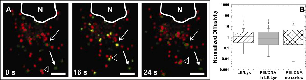
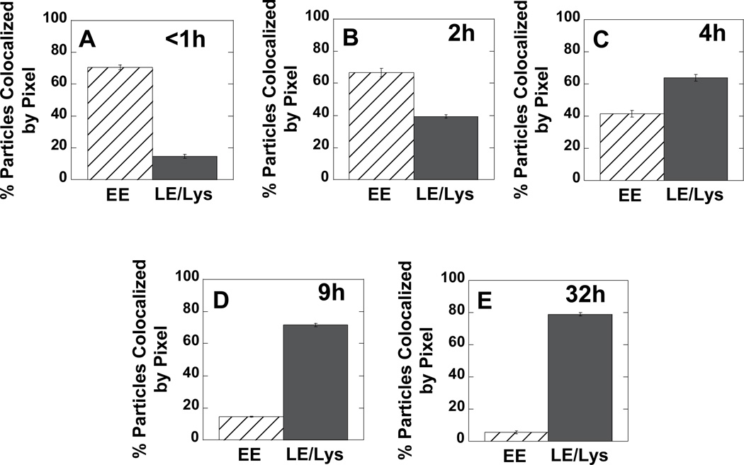
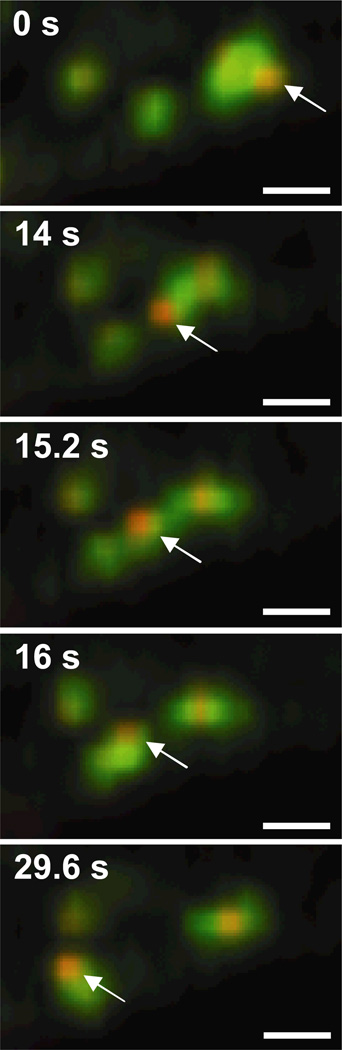
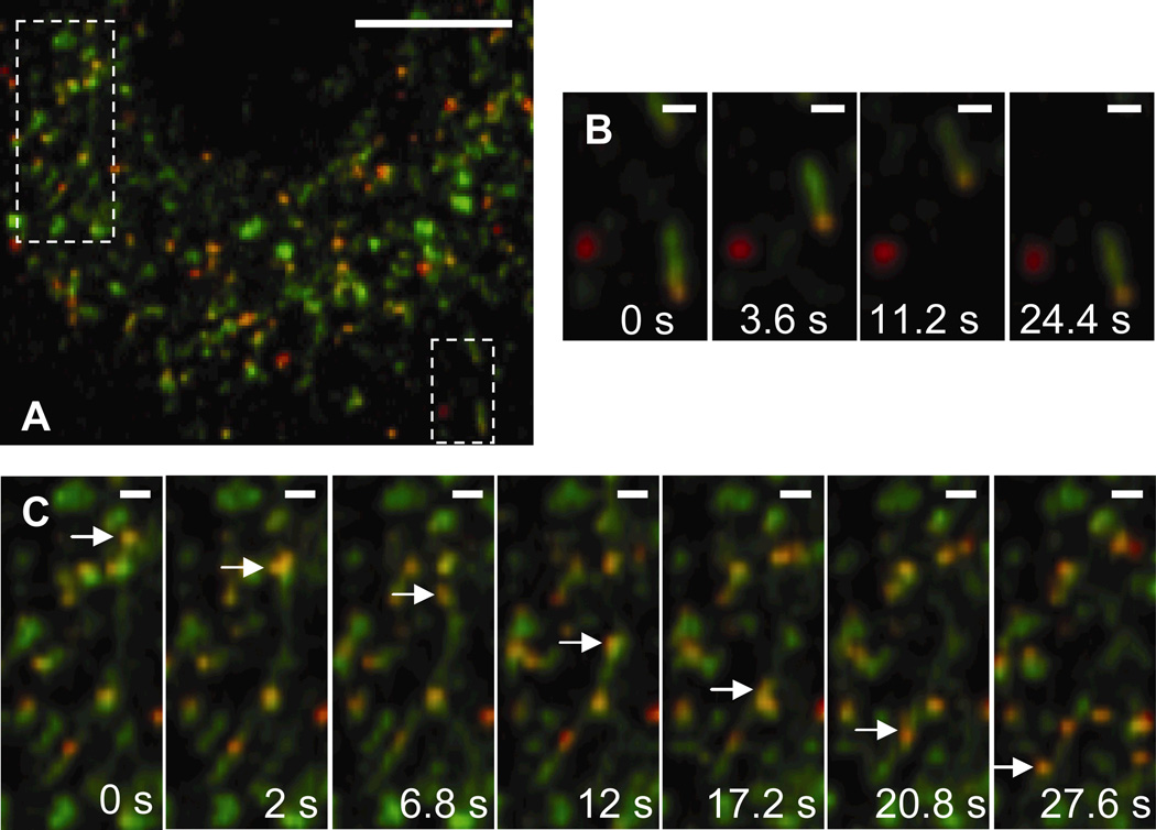
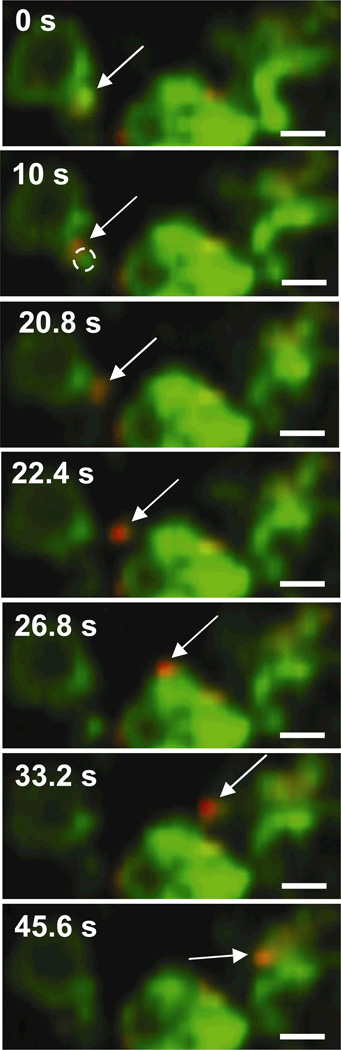
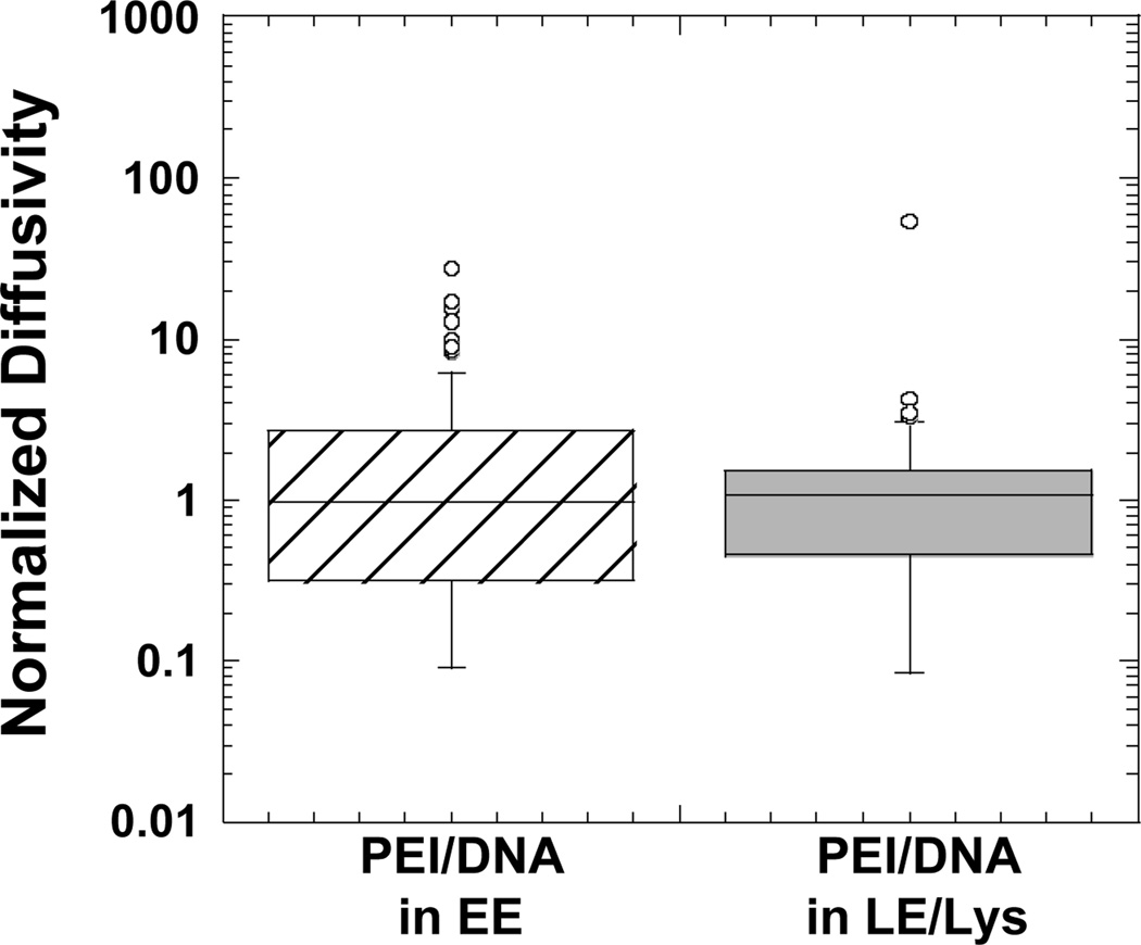
Using live-cell confocal microscopy and particle tracking technology, the simultaneous transport of intracellular vesicles of the endo-lysosomal pathway and nonviral polyethylenimine (PEI)/DNA nanocomplexes was investigated. Due to potential problems associated with the use of acid-sensitive probes in combination with a gene vector that is hypothesized to buffer the pH of intracellular vesicles, the biological location of PEI/DNA gene vectors was revealed by probing their trafficking in cells expressing fluorescent versions of either early endosome antigen 1, a protein that localizes to early endosomes, or Niemann Pick C1, a protein that localizes to late endosomes and lysosomes. Studies directly show that PEI/DNA nanoparticles are actively transported within both early and late endosomes, and display similar overall transport rates in each. Additionally, gene vector transfer between endosomes is observed. Over time post-transfection, gene vectors accumulate in late endosomes/lysosomes; however, real-time escape of vectors from membrane-bound vesicles is not observed.
Copyright © 2011 Wiley Periodicals, Inc.
Figures
Similar articles
-
Quantifying the intracellular transport of viral and nonviral gene vectors in primary neurons.Exp Biol Med (Maywood). 2007 Mar;232(3):461-9. Exp Biol Med (Maywood). 2007. PMID: 17327481
-
Niemann-Pick C1 functions independently of Niemann-Pick C2 in the initial stage of retrograde transport of membrane-impermeable lysosomal cargo.J Biol Chem. 2010 Feb 12;285(7):4983-94. doi: 10.1074/jbc.M109.037622. Epub 2009 Dec 10. J Biol Chem. 2010. PMID: 20007703 Free PMC article.
-
Exploring polyethylenimine-mediated DNA transfection and the proton sponge hypothesis.J Gene Med. 2005 May;7(5):657-63. doi: 10.1002/jgm.696. J Gene Med. 2005. PMID: 15543529
-
Endosomes, lysosomes: their implication in gene transfer.Adv Drug Deliv Rev. 2000 Mar 30;41(2):201-8. doi: 10.1016/s0169-409x(99)00066-6. Adv Drug Deliv Rev. 2000. PMID: 10699315 Review.
-
The delivery of endocytosed cargo to lysosomes.Biochem Soc Trans. 2009 Oct;37(Pt 5):1019-21. doi: 10.1042/BST0371019. Biochem Soc Trans. 2009. PMID: 19754443 Review.
Cited by
-
Vitamin D modulates human macrophage response to Mycobacterium tuberculosis DNA.Tuberculosis (Edinb). 2019 May;116S:S131-S137. doi: 10.1016/j.tube.2019.04.021. Epub 2019 May 3. Tuberculosis (Edinb). 2019. PMID: 31085128 Free PMC article.
-
Particle tracking in drug and gene delivery research: State-of-the-art applications and methods.Adv Drug Deliv Rev. 2015 Aug 30;91:70-91. doi: 10.1016/j.addr.2015.03.017. Epub 2015 Apr 7. Adv Drug Deliv Rev. 2015. PMID: 25858664 Free PMC article. Review.
-
Quantitative Particle Uptake by Cells as Analyzed by Different Methods.Angew Chem Int Ed Engl. 2020 Mar 27;59(14):5438-5453. doi: 10.1002/anie.201906303. Epub 2019 Dec 13. Angew Chem Int Ed Engl. 2020. PMID: 31657113 Free PMC article. Review.
-
IFN-stimulated metabolite transporter ENT3 facilitates viral genome release.EMBO Rep. 2023 Mar 6;24(3):e55286. doi: 10.15252/embr.202255286. Epub 2023 Jan 18. EMBO Rep. 2023. PMID: 36652307 Free PMC article.
-
Nonviral targeted mRNA delivery: principles, progresses, and challenges.MedComm (2020). 2025 Jan 2;6(1):e70035. doi: 10.1002/mco2.70035. eCollection 2025 Jan. MedComm (2020). 2025. PMID: 39760110 Free PMC article. Review.
References
-
- Bausinger R, von Gersdorff K, Braeckmans K, Ogris M, Wagner E, Zumbusch A, Brauchle C. The transport of nanosized gene carriers unraveled by live-cell imaging. Angewandte Chemie-International Edition. 2006;45(10):1568–1572. - PubMed
-
- Bieber T, Meissner W, Kostin S, Niemann A, Elsasser HP. Intracellular route and transcriptional competence of polyethylenimine-DNA complexes. Journal of Controlled Release. 2002;82(2–3):441–454. - PubMed
-
- Dauty E, Verkman AS. Actin cytoskeleton as the principal determinant of size-dependent DNA mobility in cytoplasm: a new barrier for non-viral gene delivery. J Biol Chem. 2005;280(9):7823–7828. - PubMed
-
- de Bruin K, Ruthardt N, von Gersdorff K, Bausinger R, Wagner E, Ogris M, Brauchle C. Cellular dynamics of EGF receptor-targeted synthetic viruses (vol 15, pg 1297, 2007) Molecular Therapy. 2007;15(9):1735–1735. - PubMed
Publication types
MeSH terms
Grants and funding
LinkOut - more resources
Full Text Sources
