. 2012 Mar 1;21(5):1062-77.
doi: 10.1093/hmg/ddr536. Epub 2011 Nov 17.

Functional genomic analysis unravels a metabolic-inflammatory interplay in adrenoleukodystrophy

Affiliations

Functional genomic analysis unravels a metabolic-inflammatory interplay in adrenoleukodystrophy

Agatha Schlüter et al. Hum Mol Genet. .

Abstract

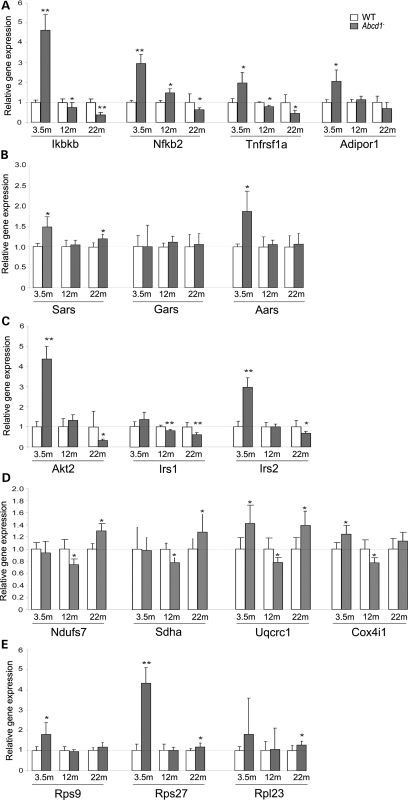
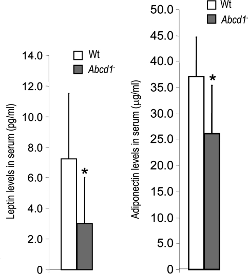
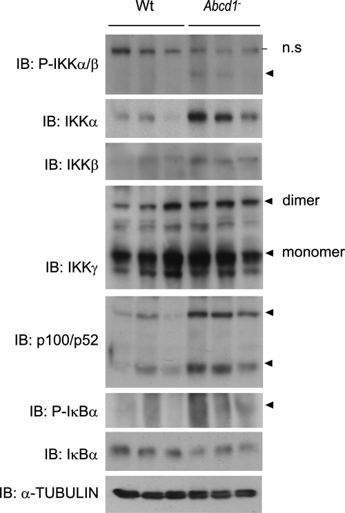
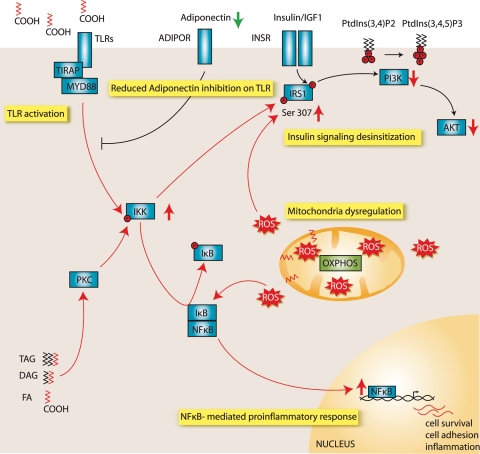
X-linked adrenoleukodystrophy (X-ALD) is an inherited disorder characterized by axonopathy and demyelination in the central nervous system and adrenal insufficiency. Main X-ALD phenotypes are: (i) an adult adrenomyeloneuropathy (AMN) with axonopathy in spinal cords, (ii) cerebral AMN with brain demyelination (cAMN) and (iii) a childhood variant, cALD, characterized by severe cerebral demyelination. Loss of function of the ABCD1 peroxisomal fatty acid transporter and subsequent accumulation of very-long-chain fatty acids (VLCFAs) are the common culprits to all forms of X-ALD, an aberrant microglial activation accounts for the cerebral forms, whereas inflammation allegedly plays no role in AMN. How VLCFA accumulation leads to neurodegeneration and what factors account for the dissimilar clinical outcomes and prognosis of X-ALD variants remain elusive. To gain insights into these questions, we undertook a transcriptomic approach followed by a functional-enrichment analysis in spinal cords of the animal model of AMN, the Abcd1(-) null mice, and in normal-appearing white matter of cAMN and cALD patients. We report that the mouse model shares with cAMN and cALD a common signature comprising dysregulation of oxidative phosphorylation, adipocytokine and insulin signaling pathways, and protein synthesis. Functional validation by quantitative polymerase chain reaction, western blots and assays in spinal cord organotypic cultures confirmed the interplay of these pathways through IkB kinase, being VLCFA in excess a causal, upstream trigger promoting the altered signature. We conclude that X-ALD is, in all its variants, a metabolic/inflammatory syndrome, which may offer new targets in X-ALD therapeutics.


Figures

Figure 1.
Figure 1.
(A) A procedural diagram that indicates the pipeline followed to obtain the disease signature and progression in X-ALD mice and patients. (B) A hierarchical clustering heat-map of the expression intensities of the significantly regulated transcripts (on the Y-axis) versus the X-ALD patients and controls arrays (on the X-axis). Interestingly, we observed a clear separation between X-ALD patients and controls suggesting that hX-ALD pathology is concomitant to an alteration of transcriptional profiles.
Figure 2.
Figure 2.
Common dysregulated pathways in spinal cords of ALD mice at 3.5, 12 and 22 months, and cerebral white matter of child and adult ALD patients. Significant KEGG pathways were identified by using the statistics gene set enrichment analysis (GSEA) (G) and hypergeometric distribution function (H). Only the pathways statistically significant in at least two tests are represented. Black spots represent dysregulated pathway when probability value P is <0.05 according to the H-test, which detects changes but does not inform about their up or down direction. Red and green spots represent up- and downregulated pathway expression based on the G-test when probability value of P is <0.05, orange spots means dysregulated pathway based on the G-test with a probability value of <0.05, in case the gene set does not have a clear tendency to be up- or downregulated. Blue circle means significant pathway with a probability value of P< 0.0001.
Figure 3.
Figure 3.
Validation of dysregulated pathways at three different ages in Abcd1 mice spinal cords. qRT-PCR measurements of (A) adipocytokine signaling pathway, (B) aminoacyl-tRNA biosynthesis, (C) insulin signaling pathway, (D) oxidative phosphorylation and (E) ribosomal subunits. Ikbkb, NF-κB2, Tnfrsf1a, Adipor1, Sars, Gars, Aars, Akt2, Irs1, Irs2, Ndufs7, Sdha, Uqcrc1, Cox4i1, Rps9, Rps27 and Rpl23 mRNA has been quantified by qRT-PCR. 18S was used as an internal control. Significant differences have been determined by Student's t-test. Significant differences are shown as *P< 0.05 and **P< 0.01.
Figure 4.
Figure 4.
Leptin concentration was lower in serum from Abcd1 mice (A). Leptin (pg/ml) was measured in serum from 12-month-old Wt (n= 8) and Abcd1 (n= 9) mice. Adiponectin concentration was also lower in serum from Abcd1 mice (B). Adiponectin (µg/ml) was measured in serum from 12-month-old Wt (n= 10) and Abcd1 mice (n= 12). Statistical analysis was done by Student's t-test: *P< 0.05.
Figure 5.
Figure 5.
(A) Insulin signaling pathway in Wt and Abcd1 mouse spinal cords at 12 months of age. Western blots to monitor contents of IRS1, p-IRS1 Ser307, AKT, p-AKT Thr308, Ser473, p-S6K Thr389, JNK and p-JNK Thr183 and Tyr185 have been performed in whole spinal cord lysates from Wt and Abcd1 mice (n = 6 individuals per genotype). Representative blots are shown. Statistical analysis was done by Student's t-test: *P < 0.05. The phosphorylation of IRS1 in Ser307 was induced in Abcd1 mice spinal cord in the absence of insulin signaling activation, as shown by the lack of activation of kinases pS6K and JNK. (B) Phosphorylation of IRS1 ser307 in organotypic spinal cord slice culture (OSCSC). Western blots to monitor phosphorylation levels of IRS1 ser 307 have been performed on whole protein extracts from OSCSC (n = 6 cultures per genotype, Abcd1 or Wt littermates) incubated 24 h with BSA-conjugated C26:0 50 µm or BSA alone as control. Representative western blots are shown. C26:0 induces the IRS1 Ser307 phosphorylation in Wt but not Abcd1-derived OSCSC.
Figure 6.
Figure 6.
Activation of kinases IKK in mouse spinal cord (SC) at 12 months of age. Western blots to monitor levels of IKKα, IKKβ, IKKγ, NF-κB2 (p100/p52) and IκBα were performed in whole spinal cord lysates from Wt and Abcd1 mice (n= 6 individuals per genotype). Activated IKKs phosphorylate IκBα that liberates the NF-κB complex, which subsequently translocates into the nucleus.
Figure 7.
Figure 7.
Activation of TLR/NF-κB in mouse spinal cords at 12 months of age. A q-PCR was performed to measure a gene array including TLRs, TLR adaptors, NF-κB forms, several cytokines and their receptors. Only differentially expressed genes are shown (n= 6 individuals per genotype). The data point to a general activation of TLR/NF-κB pathways, wherein we highlight the upregulation of Tlr8, Tlr5, Nfkbib, RANTES (Ccl5), Ccr1, Ccr2, Ccr7, Il1b, Tnf and Lta. Significant differences have been determined by Student's t-test: *P< 0.05, **P< 0.01 and ***P< 0.001.
Figure 8.
Figure 8.
The role of oxidative stress in ALD progression in spinal cord is already manifesting as early as 3.5 months of age. The picture shows dysregulated gene expression, induced (red triangle) and repressed (green triangle), of processes and components involved in ROS generation (black stars) and antioxidant defense (white stars). Perturbations in oxidative phosphorylation, iron and calcium homeostasis and changes in GSH homeostasis may altogether represent causes and consequences of increased oxidative stress. ROS might activate NF-κB leading to the regulation of genes involved in cell survival and inflammation. 12-Hydroxyeicosatetrenoate (12-HETE), arachidonate 12-lipoxygenase (ALOX12), ATPase, Ca2+ transporting, cardiac muscle, slow twitch 2 (ATP2A2), ATPase, Ca2+ transporting, plasma membrane 1 (ATP2B1), calcium-channel, voltage-dependent, alpha 2/delta subunit 3 (CACNA2D3), calcium-channel, voltage-dependent, gamma subunit 5 (CACNG5), calcium-channel, voltage-dependent, L-type, alpha 1D subunit (CACNA1D), calcium/calmodulin-dependent protein kinase (CaM kinase) II delta (CAMK2D), calmodulin 1 (CALM1), protein phosphatase 3, catalytic subunit, alpha isoform (PPP3CA), ceruloplasmin (CP), coenzyme Q10 (CQ), coproporphyrinogen oxidase (CPOX), cystathionine beta-synthase (CBS), cytochrome b-245 (CYBA), cytochrome c (CytC), DLST, dihydroxyacetone-phosphate (DHAP), electron transport chain complexes I:V (C-I:C-V), epoxide hydrolase 1, microsomal (EPHX1), glucose-6-phosphate dehydrogenase X-linked (G6PDH), GCLC, GRX, GSH, glutathione disulfide (GSSG), GPX, glutathione reductase (GSR), GST, GSS, glyceraldehyde-3-phosphate (G3P), glycerol-3-phosphate dehydrogenase (GPDH), glyoxalase 1 (GLO1), glyoxylate reductase/hydroxypyruvate reductase (GRHPR), nuclear factor of kappa light polypeptide gene enhancer in B-cells inhibitor alpha (NFKBIA or IκBα), conserved helix–loop–helix ubiquitous kinase (CHUK or IKKα), inhibitor of kappa light polypeptide gene enhancer in B-cells, kinase beta (IKBKB or IKKβ), inhibitor of kappa light polypeptide gene enhancer in B-cells, kinase gamma (IKBKG or IKKγ), lipid hydroperoxide (LOOH), lipid hydroxide (LOH), malic enzyme (ME), NADPH oxidase 1 (NOX1), nuclear factor of kappa light polypeptide gene enhancer in B-cells 1 and 2 (NF-κB1 and NF-κB2), OGDH, PRX, phosphodiesterase 1A, calmodulin-dependent (PDE1A), PGPX, phosphorylase kinase, alpha 1 (PHKA1), pyruvate dehydrogenase (PDH), solute carrier family 8 (sodium/calcium exchanger), member 1 (SLC8A1), thioredoxin (TRX), TCA, triosephosphate isomerase 1 (TPI), uncoupling protein 2 (UCP2), uroporphyrinogen decarboxylase (UROD), uroporphyrinogen III synthase (UROS).
Figure 9.
Figure 9.
Proposed model for X-ALD disease mechanism. Taking together our functional validation that ensued our transcriptomic data mining and the experimental evidence provided, we put forth the following model: disease progression appears mediated by the successive action of a primarily VLCFA excess, mitochondrial dysregulation combined with ROS production and antioxidant defenses depletion, insulin desensitization, TLR activation and NF-κB-mediated proinflammatory response. The increased lipid load induces a stress response leading to the production of ROS contributing all together to a mitochondrial dysregulation. The activation of IKKs links VLCFA-induced metabolic deficiency with an NF-κB-mediated proinflammatory response. IKK activation desensitizes insulin signaling via phosphorylation of IRS1 on serines. Insulin controls glucose uptake through tyrosine phosphorylation of insulin receptor substrate (IRS) proteins. Activation of phosphatidylinositol 3-kinase (PI3K) and generation of phosphatidylinositol-3,4,5-trisphosphate (PtdIns(3,4,5)P3) activates AKT. But IRS1 serine phosphorylation attenuates insulin sensitivity and subsequently decreases glucose uptake and insulin-induced metabolism. IKKs can be induced by FA via the activation of TLRs and PKC, and the latter can be activated by DAG. The low levels of adiponectin might contribute to the increased proinflammatory response in X-ALD by reducing the inhibitory adiponectin effect on TLR signaling.

References

    1. Ferrer I., Aubourg P., Pujol A. General aspects and neuropathology of X-linked adrenoleukodystrophy. Brain Pathol. 2010;20:817–830. doi:10.1111/j.1750-3639.2010.00390.x. - DOI - PMC - PubMed
    1. Mosser J., Douar A.M., Sarde C.O., Kioschis P., Feil R., Moser H., Poustka A.M., Mandel J.L., Aubourg P. Putative X-linked adrenoleukodystrophy gene shares unexpected homology with ABC transporters. Nature. 1993;361:726–730. doi:10.1038/361726a0. - DOI - PubMed
    1. van Roermund C.W., Visser W.F., Ijlst L., van Cruchten A., Boek M., Kulik W., Waterham H.R., Wanders R.J. The human peroxisomal ABC half transporter ALDP functions as a homodimer and accepts acyl-CoA esters. FASEB J. 2008;22:4201–4208. doi:10.1096/fj.08-110866. - DOI - PubMed
    1. Kemp S., Pujol A., Waterham H.R., van Geel B.M., Boehm C.D., Raymond G.V., Cutting G.R., Wanders R.J., Moser H.W. ABCD1 mutations and the X-linked adrenoleukodystrophy mutation database: role in diagnosis and clinical correlations. Hum. Mutat. 2001;18:499–515. doi:10.1002/humu.1227. - DOI - PubMed
    1. Moser H., Smith K.D., Watkins P.A., Powers J., Moser A.B. In: The Metabolic and Molecular Bases of Inherited Disease. Scriver C., editor. New York: McGraw-Hill; 2001. pp. 3257–3301.

Publication types

MeSH terms