

Blocking IL-1 signaling rescues cognition, attenuates tau pathology, and restores neuronal β-catenin pathway function in an Alzheimer's disease model
- PMID: 22095718
- PMCID: PMC4072218
- DOI: 10.4049/jimmunol.1100620
Blocking IL-1 signaling rescues cognition, attenuates tau pathology, and restores neuronal β-catenin pathway function in an Alzheimer's disease model
Abstract
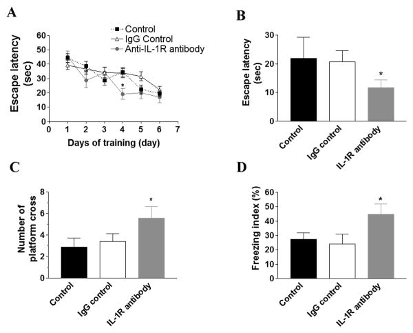
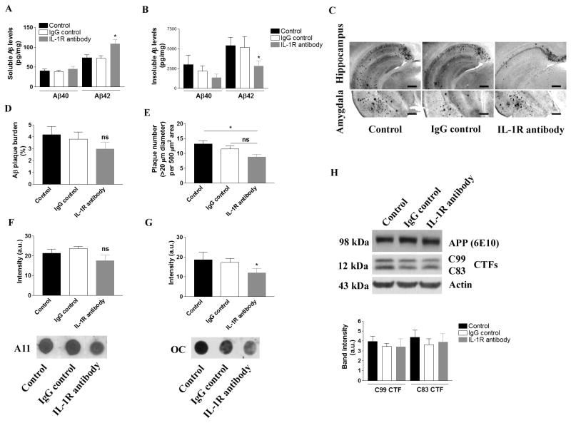
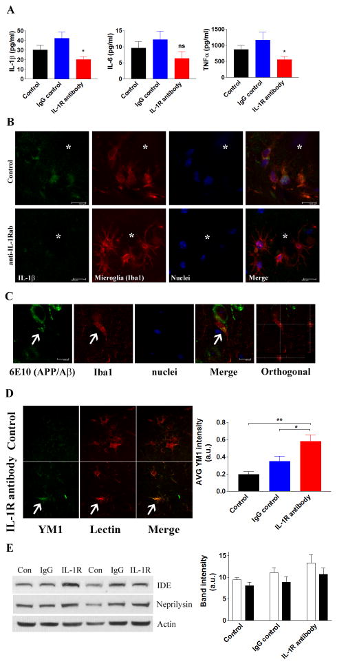
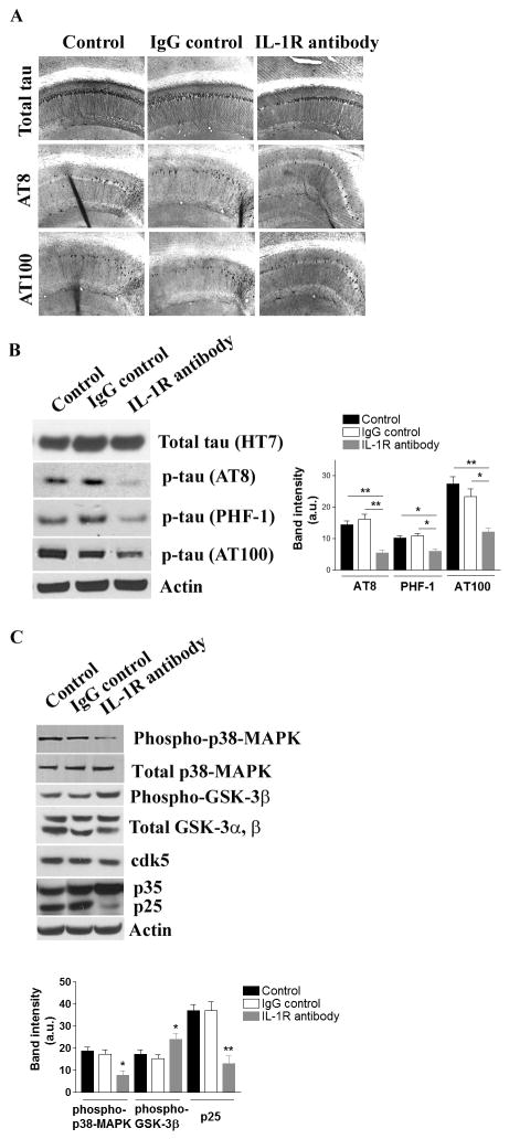
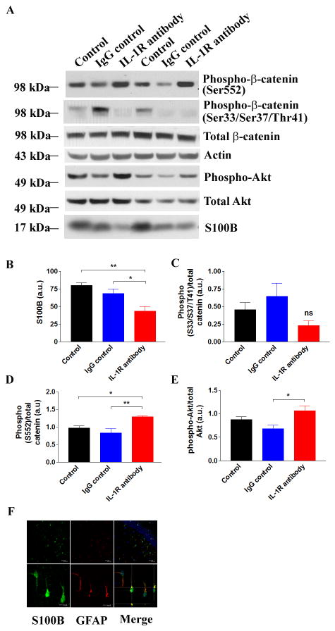
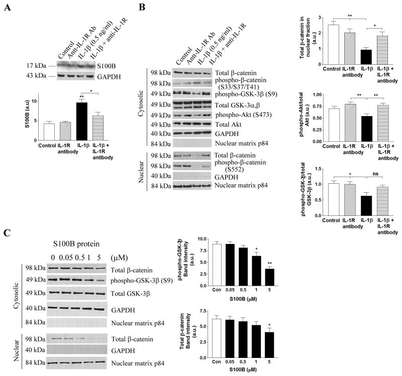
Inflammation is a key pathological hallmark of Alzheimer's disease (AD), although its impact on disease progression and neurodegeneration remains an area of active investigation. Among numerous inflammatory cytokines associated with AD, IL-1β in particular has been implicated in playing a pathogenic role. In this study, we sought to investigate whether inhibition of IL-1β signaling provides disease-modifying benefits in an AD mouse model and, if so, by what molecular mechanisms. We report that chronic dosing of 3xTg-AD mice with an IL-1R blocking Ab significantly alters brain inflammatory responses, alleviates cognitive deficits, markedly attenuates tau pathology, and partly reduces certain fibrillar and oligomeric forms of amyloid-β. Alterations in inflammatory responses correspond to reduced NF-κB activity. Furthermore, inhibition of IL-1 signaling reduces the activity of several tau kinases in the brain, including cdk5/p25, GSK-3β, and p38-MAPK, and also reduces phosphorylated tau levels. We also detected a reduction in the astrocyte-derived cytokine, S100B, and in the extent of neuronal Wnt/β-catenin signaling in 3xTg-AD brains, and provided in vitro evidence that these changes may, in part, provide a mechanistic link between IL-1 signaling and GSK-3β activation. Taken together, our results suggest that the IL-1 signaling cascade may be involved in one of the key disease mechanisms for AD.
Figures
Similar articles
-
Neuronal deficiency of p38α-MAPK ameliorates symptoms and pathology of APP or Tau-transgenic Alzheimer's mouse models.FASEB J. 2020 Jul;34(7):9628-9649. doi: 10.1096/fj.201902731RR. Epub 2020 May 31. FASEB J. 2020. PMID: 32475008
-
Restoration of lipoxin A4 signaling reduces Alzheimer's disease-like pathology in the 3xTg-AD mouse model.J Alzheimers Dis. 2015;43(3):893-903. doi: 10.3233/JAD-141335. J Alzheimers Dis. 2015. PMID: 25125468 Free PMC article.
-
Selenomethionine Mitigates Cognitive Decline by Targeting Both Tau Hyperphosphorylation and Autophagic Clearance in an Alzheimer's Disease Mouse Model.J Neurosci. 2017 Mar 1;37(9):2449-2462. doi: 10.1523/JNEUROSCI.3229-16.2017. Epub 2017 Jan 30. J Neurosci. 2017. PMID: 28137967 Free PMC article.
-
Effects of CX3CR1 and Fractalkine Chemokines in Amyloid Beta Clearance and p-Tau Accumulation in Alzheimer's Disease (AD) Rodent Models: Is Fractalkine a Systemic Biomarker for AD?Curr Alzheimer Res. 2016;13(4):403-12. doi: 10.2174/1567205013666151116125714. Curr Alzheimer Res. 2016. PMID: 26567742 Review.
-
Roles of Cytokines in Alzheimer's Disease.Int J Mol Sci. 2024 May 26;25(11):5803. doi: 10.3390/ijms25115803. Int J Mol Sci. 2024. PMID: 38891990 Free PMC article. Review.
Cited by
-
Suppression of TLR signaling by targeting TIR domain-containing proteins.Curr Protein Pept Sci. 2012 Dec;13(8):776-88. doi: 10.2174/138920312804871148. Curr Protein Pept Sci. 2012. PMID: 23305364 Free PMC article. Review.
-
Cochlear inflammaging: cellular and molecular players of the innate and adaptive immune system in age-related hearing loss.Front Neurol. 2023 Nov 22;14:1308823. doi: 10.3389/fneur.2023.1308823. eCollection 2023. Front Neurol. 2023. PMID: 38073631 Free PMC article. Review.
-
Interactions between inflammation, sex steroids, and Alzheimer's disease risk factors.Front Neuroendocrinol. 2016 Oct;43:60-82. doi: 10.1016/j.yfrne.2016.09.001. Epub 2016 Sep 17. Front Neuroendocrinol. 2016. PMID: 27651175 Free PMC article. Review.
-
Targeting Microglia in Alzheimer's Disease: Pathogenesis and Potential Therapeutic Strategies.Biomolecules. 2024 Jul 11;14(7):833. doi: 10.3390/biom14070833. Biomolecules. 2024. PMID: 39062547 Free PMC article. Review.
-
The Dichotomous Role of Inflammation in the CNS: A Mitochondrial Point of View.Biomolecules. 2020 Oct 13;10(10):1437. doi: 10.3390/biom10101437. Biomolecules. 2020. PMID: 33066071 Free PMC article. Review.
References
-
- Lee YJ, Han SB, Nam SY, Oh KW, Hong JT. Inflammation and Alzheimer's disease. Archives of pharmacal research. 2010;33:1539–1556. - PubMed
-
- McGeer PL, Schulzer M, McGeer EG. Arthritis and anti-inflammatory agents as possible protective factors for Alzheimer's disease: a review of 17 epidemiologic studies. Neurology. 1996;47:425–432. - PubMed
-
- Breitner JC, Gau BA, Welsh KA, Plassman BL, McDonald WM, Helms MJ, Anthony JC. Inverse association of anti-inflammatory treatments and Alzheimer's disease: initial results of a co-twin control study. Neurology. 1994;44:227–232. - PubMed
Publication types
MeSH terms
Substances
Grants and funding
- P01 AG000538/AG/NIA NIH HHS/United States
- NS050895/NS/NINDS NIH HHS/United States
- RF1 AG020241/AG/NIA NIH HHS/United States
- R01 NS050895/NS/NINDS NIH HHS/United States
- AG00538/AG/NIA NIH HHS/United States
- R01AG20335/AG/NIA NIH HHS/United States
- P50AG16573/AG/NIA NIH HHS/United States
- R01 AG020241/AG/NIA NIH HHS/United States
- R01 AG021982/AG/NIA NIH HHS/United States
- AG020241/AG/NIA NIH HHS/United States
- R01 AG020335/AG/NIA NIH HHS/United States
- K99AR054695/AR/NIAMS NIH HHS/United States
- K99 AR054695/AR/NIAMS NIH HHS/United States
- P50 AG016573/AG/NIA NIH HHS/United States
LinkOut - more resources
Full Text Sources
Other Literature Sources
Medical
Molecular Biology Databases
Miscellaneous
