Review
. 2011 Dec;3(12):701-12.
doi: 10.1002/emmm.201100175. Epub 2011 Sep 23.

Regeneration of the heart

Affiliations
Review

Regeneration of the heart

Matthew L Steinhauser et al. EMBO Mol Med. 2011 Dec.

Abstract

The death of cardiac myocytes diminishes the heart's pump function and is a major cause of heart failure, one of the dominant causes of death worldwide. Other than transplantation, there are no therapies that directly address the loss of cardiac myocytes, which explains the current excitement in cardiac regeneration. The field is evolving in two important directions. First, although endogenous mammalian cardiac regeneration clearly seems to decline rapidly after birth, it may still persist in adulthood. The careful elucidation of the cellular and molecular mechanisms of endogenous heart regeneration may therefore provide an opportunity for developing therapeutic interventions that amplify this process. Second, recent breakthroughs have enabled reprogramming of cells that were apparently terminally differentiated, either by dedifferentiation into pluripotent stem cells or by transdifferentiation into cardiac myocytes. These achievements challenge our conceptions of what is possible in terms of heart regeneration. In this review, we discuss the current status of research on cardiac regeneration, with a focus on the challenges that hold back therapeutic development.


Figures

Figure 1
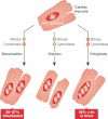
Figure 1. The majority of post-natal human DNA synthesis in the heart does not lead to new myocyte formation
Cardiac myocytes can complete S-phase, followed by mitosis and cytokinesis (centre) resulting in myocyte doubling. Cardiac myocytes can also complete mitosis without cytokinesis (left), resulting in a binucleated cell. Cardiac myocytes can also undergo chromosomal replication without completing either mitosis or cytokinesis (right), resulting in polyploidy nuclei. By the completion of post-natal development, the majority of human myocyte nuclei contain ≥4n chromosomal copies.
Figure 2
Figure 2. Lineage-mapping in the adult heart
Left: Theoretical progenitor lineage-mapping is depicted. Progenitors would be selectively marked by fluorescent protein expression. After injury, the appearance of fluorescently labelled cardiac myocytes would support the concept that these progenitors were contributing to new myocyte formation. Right: Differentiated cell (cardiac myocyte) lineage-mapping. Upon treatment of the MerCreMer-ZEG mouse with OH-tamoxifen, approximately 80% of the cardiac myocytes undergo a permanent switch from β-galactosidase to GFP expression. The dilution of the GFP+ cardiac myocyte pool after injury is consistent with repletion by β-gal+ progenitors.
Figure 3
Figure 3. Possible recapitulation of developmental paradigms by endogenous post-natal cardiac stem cells
Between mesodermal development and the emergence of cardiac myocytes, cardiovascular progenitors express a number of markers that have also been detected in the various post-natal cardiac stem cell (CSC) preparations. Expression as measured by messenger RNA (mRNA) or protein expression is denoted with (+). Absent expression is denoted by (−). Blank = untested.
Figure 4
Figure 4. The challenge of regenerating the heart
Both exogenously delivered cell therapies and progenitors in the endogenous niche encounter a similar hostile environment after myocardial injury, often including inadequate blood supply (ischemia), inflammation and fibrosis/scarring. Regenerative pathways may be activated by as yet unknown paracrine pathways, responsible for recruiting progenitors from the niche, stimulating proliferation and coaxing differentiation.
Figure 5
Figure 5. Proposed mechanisms of action for cell-based therapies
In theory, exogenously delivered cells may directly differentiate into endothelial cells, smooth muscle cells and cardiac myocytes. They may also release paracrine factors which may result in non-regenerative effects, such as immunomodulation, angiogenesis or cardioprotection. Recent work from our laboratory suggests that a dominant mechanism achieved with bone marrow progenitor therapy may be via the activation of endogenous progenitor recruitment (Loffredo et al, 2011).

Similar articles

Cited by

References

    1. Adler CP, Friedburg H. Myocardial DNA content, ploidy level and cell number in geriatric hearts: post-mortem examinations of human myocardium in old age. J Mol Cell Cardiol. 1986;18:39–53. - PubMed
    1. Amariglio N, Hirshberg A, Scheithauer BW, Cohen Y, Loewenthal R, Trakhtenbrot L, Paz N, Koren-Michowitz M, Waldman D, Leider-Trejo L, et al. Donor-derived brain tumor following neural stem cell transplantation in an ataxia telangiectasia patient. PLoS Med. 2009;6:e1000029. - PMC - PubMed
    1. Andersen DC, Andersen P, Schneider M, Jensen HB, Sheikh SP. Murine “cardiospheres” are not a source of stem cells with cardiomyogenic potential. Stem Cells. 2009;27:1571–1581. - PubMed
    1. Astorri E, Chizzola A, Visioli O, Anversa P, Olivetti G, Vitali-Mazza L. Right ventricular hypertrophy-a cytometric study on 55 human hearts. J Mol Cell Cardiol. 1971;2:99–110. - PubMed
    1. Balsam LB, Wagers AJ, Christensen JL, Kofidis T, Weissman IL, Robbins RC. Haematopoietic stem cells adopt mature haematopoietic fates in ischaemic myocardium. Nature. 2004;428:668–673. - PubMed