. 2012 Feb;32(2):397-405.
doi: 10.1161/ATVBAHA.111.239897. Epub 2011 Nov 17.

Regulation of peroxisome proliferator-activated receptor-γ by angiotensin II via transforming growth factor-β1-activated p38 mitogen-activated protein kinase in aortic smooth muscle cells

Affiliations

Regulation of peroxisome proliferator-activated receptor-γ by angiotensin II via transforming growth factor-β1-activated p38 mitogen-activated protein kinase in aortic smooth muscle cells

Venkateswaran Subramanian et al. Arterioscler Thromb Vasc Biol. 2012 Feb.

Abstract

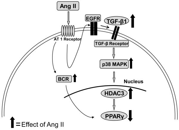
Objective: Peroxisome proliferator-activated receptor-γ (PPARγ) ligands attenuate angiotensin II (Ang II)-induced atherosclerosis through interactions with vascular smooth muscle cell (VSMC)-specific PPARγ in hypercholesterolemic mice. Therefore, the purpose of this study was to determine the mechanism of Ang II-mediated intracellular regulation of PPARγ in VSMCs.

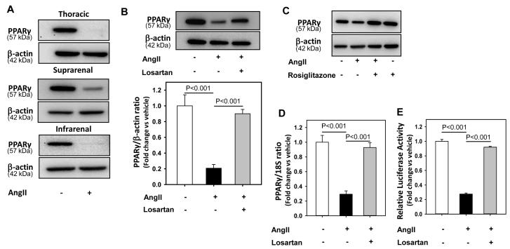
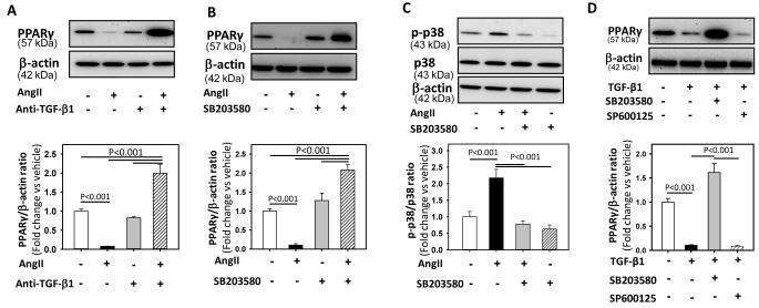
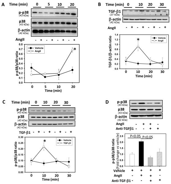
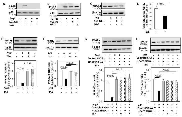
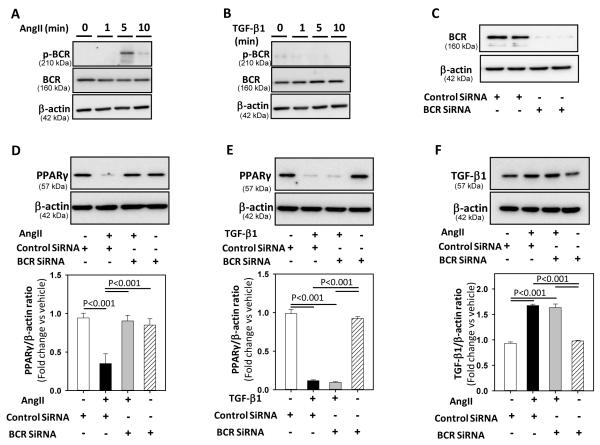
Methods and results: Incubation of cultured mouse aortic VSMCs with Ang II for 24 hours reduced abundance of PPARγ protein, mRNA, and transcriptional activity (P<0.001). This effect was attenuated by an angiotensin type 1 receptor antagonist, losartan. Ang II-induced PPARγ reduction was dependent on stimulation of transforming growth factor (TGF)-β1 as demonstrated using either a neutralizing antibody or small interfering RNA (siRNA). Ang II-induced TGF-β1 secretion was dependent on epidermal growth factor receptor kinase activation through reactive oxygen species production. Inhibition of p38 mitogen-activated protein kinase by SB203580 or siRNA inhibited both Ang II- and TGF-β1-induced PPARγ reduction. Blockade of TGF-β1 decreased p38 phosphorylation induced by Ang II. siRNA-mediated inhibition of histone deacetylase 3 attenuated p38-mediated reductions in PPARγ abundance.

Conclusions: These findings suggest that Ang II decreases PPARγ abundance in cultured VSMCs via an angiotensin type 1 receptor-dependent secretion of TGF-β1 via phosphorylation of p38 mitogen-activated protein kinase and histone deacetylase 3.


Figures

Figure 1
Figure 1. AngII decreased PPARγ protein, mRNA abundance and transcriptional activity
A: Thoracic, suprarenal, and infrarenal aorta-derived VSMCs were incubated with either vehicle (saline) or AngII (1 μM) for 24 hours, lysed, and proteins were resolved by SDS-PAGE. Western analyses were performed to detect PPARγ protein (n = 3). B: VSMCs were incubated with either vehicle (saline), AngII (1 μM) or AngII + losartan (10 μM) for 24 hours. Total cell lysates were analyzed by Western blotting using antibodies to PPARγ (n = 5). C: VSMCs were pre-incubated with either vehicle (DMSO) or rosiglitazone (10 μM) for 24 hours followed by incubation with either saline or AngII (1 μM) for 24 hours. Total cell lysates were analyzed by Western blotting using antibodies to PPARγ (n = 3). Abundance of β-actin was used as an internal control. D: VSMCs were incubated with either vehicle (saline), AngII (1 μM) or AngII + losartan (10 μM) for 24 hours. Total RNA was extracted from these cells and then subjected to qRT-PCR analyses. 18S was used as an internal control (n = 4). E: VSMCs were transiently transfected with PPARγ promoter luciferase reporter construct along with plasmid encoding renilla luciferase. After transfection, cells were serum-deprived and incubated with either vehicle (saline) or AngII for 24 hours. Transfection efficiency was adjusted by normalizing firefly luciferase activities to renilla luciferase activities (n = 4). Results are represented as means ± SEMs; Data were analyzed by one-way ANOVA with a Holm-Sidak multiple comparison post-hoc test.
Figure 2
Figure 2. AngII decreased PPARγ via the activation of TGF-β1 and p38 MAPK
A: VSMCs were incubated with either vehicle (saline), AngII (1 μM) or AngII + anti-TGF-β1 antibody (2 μg/ml) for 24 hours. Total cell lysates were analyzed by Western blotting using antibodies to PPARγ (n = 4). B: VSMCs were incubated with vehicle (DMSO), AngII (1 μM) or AngII + SB-203580 (10 μM) for 24 hours. Total cell lysates were analyzed by Western blotting using antibodies to PPARγ (n = 4). C: VSMCs were pre-incubated with SB-203580 (10 μM for 30 minutes) followed by AngII incubation for 30 minutes. Total cell lysates were analyzed by Western blot using antibodies to phospho p38. D: VSMCs were incubated with vehicle (DMSO), TGF-β1 (10 ng/ml) or TGF-β1 + SB-203580 (10 μM) / SP600125 (10 μM) for 24 hours. Total cell lysates were analyzed by Western blotting using antibodies to PPARγ. β-actin was used as an internal control (n = 4). Results are represented as means ± SEMs; Data were analyzed by Student’s t test or one-way ANOVA with a Holm-Sidak multiple comparison post-hoc test.
Figure 3
Figure 3. AngII increased p38 phosphorylation and TGF-β1 expression
A: VSMCs were incubated with AngII (1 μM) for selected time intervals (0 - 20 minutes). Total cell lysates were analyzed by Western blot using antibodies to phospho-p38 or p38. * Denotes P < 0.05 comparing AngII versus vehicle (n = 3). B: VSMCs were incubated with AngII (1 μM) for selected time intervals (0 - 30 minutes). Total cell lysates were analyzed by Western blotting using a TGF-β1 antibody. β-actin was used as an internal control. * Denotes P < 0.05 comparing AngII versus vehicle and TGF-β1 antibody + AngII (n = 3). Results are represented as means ± SEMs. C: VSMCs were incubated with vehicle or TGF-β1 (10 ng/ml) for selected intervals (0 - 30 minutes). Total cell lysates were analyzed by Western blotting using antibodies to phospho-p38 or p38. * Denotes P < 0.05 for TGF-β1 vs vehicle (n = 3). Results are represented as means ± SEMs; Statistical significances between vehicle and AngII / TGF-β1 at different intervals were analyzed by Student’s t-test. D: VSMCs were incubated with either vehicle (saline), AngII (1 μM) or AngII + anti-TGF-β1 antibody (2 μg/ml) for 30 minutes. Total cell lysates were analyzed by Western blot using antibodies to phospho-p38 or p38 (n = 3). Results are represented as means ± SEMs; Data were analyzed by one-way ANOVA with a Holm-Sidak multiple comparison post-hoc test.
Figure 4
Figure 4. P38 MAPK mediated AngII-induced reduction in PPARγ via HDAC-3
A, B: VSMCs were pre-incubated with AG1478 (100 nM), or NAC (10 mM) for 30 minutes followed by incubation with either vehicle (saline) or AngII (A) / recombinant TGF-β1 (B) for 20 and 10 minutes, respectively. Total cell lysates were analyzed by Western blot using antibodies to phospho-p38 or p38 (n = 3). C: VSMCs were pre-incubated with AG1478 (100 nM), or NAC (10 mM) for 30 minutes followed by incubation with either vehicle (saline) or AngII for 10 minutes. Total cell lysates were analyzed by Western blot using antibodies to TGF-β1 (n = 3). D: VSMCs were transiently transfected with a PPARγ promoter luciferase reporter construct along with plasmid encoding renilla luciferase. After transfection, cells were serum deprived and stimulated with vehicle (saline) or recombinant active p38 (100 nM) for 24 hours. Transfection efficiency was adjusted by normalizing firefly luciferase activities to renilla luciferase activities (n = 4). E, F: VSMCs were pre-incubated with TSA (1 μM) for 30 minutes followed by incubation with either vehicle (saline) or AngII (E) / recombinant active p38 (F) for 24 hours. Total cell lysates were analyzed by Western blot using antibodies to PPARγ (n = 4). G, H: VSMCs were transfected with either HDAC3 SiRNA for 48 hours or pre-incubated with TSA for 30 minutes, followed by incubation with either vehicle (saline) or AngII (G) / recombinant active p38 (H) for 24 hours. Total cell lysates were analyzed by Western blot using antibodies to PPARγ (n = 4). β-actin was used as an internal control. Results are represented as means ± SEMs; Data were analyzed by one-way ANOVA with a Holm-Sidak multiple comparison post-hoc test.
Figure 5
Figure 5. BCR-kinase silencing prevented AngII, but not TGF-β1-induced downregulation of PPARγ
A, B: VSMCs were incubated with vehicle or AngII (A) / TGF-β1 (B) for selected intervals (0 - 10 minutes). Total cell lysates were analyzed by Western blotting using antibodies to phospho-BCR kinase or BCR kinase (n=3). C: VSMCs were transfected with either control, or BCR kinase siRNA for 48 hours and total cell lysates were analyzed by Western blot using antibodies to BCR kinase (n = 3). D, E: VSMCs were transfected with either control or BCR kinase SiRNA for 48 hours, followed by incubation with either vehicle (DMSO) or AngII (D) / recombinant TGF-β1 (E) for 24 hours. Total cell lysates were analyzed by Western blot using antibodies to PPARγ (n = 3-4). F: VSMCs were transfected with either control or BCR kinase siRNA for 48 hours, followed by incubation with either vehicle (saline) or AngII for 10 minutes. Total cell lysates were analyzed by Western blot using antibodies to TGF-β1 (n = 3). β-actin was used as an internal control. Results are represented as means ± SEMs; Data were analyzed by one-way ANOVA with a Holm-Sidak multiple comparison post-hoc test.
Figure 6
Figure 6. Proposed schematic representation of signaling pathways involved in the regulation of AngII induced decreases in PPARγ in aortic VSMCs
AngII-induced decrease in PPARγ protein was regulated via AT1 receptor activation of TGF-β1 leading to subsequent induction of p38 MAPK mediated HDAC3.

References

    1. Schoonjans K, Martin G, Staels B, Auwerx J. Peroxisome proliferator-activated receptors, orphans with ligands and functions. Current Opinion in Lipidol. 1997;8:159–166. - PubMed
    1. Olefsky JM. Treatment of insulin resistance with peroxisome proliferator-activated receptor gamma agonists. J Clin Invest. 2000;106:467–472. - PMC - PubMed
    1. Hsueh WA, Bruemmer D. Peroxisome proliferator-activated receptor gamma: implications for cardiovascular disease. Hypertension. 2004;43:297–305. - PubMed
    1. Levi Z, Shaish A, Yacov N, Levkovitz H, Trestman S, Gerber Y, Cohen H, Dvir A, Rhachmani R, Ravid M, Harats D. Rosiglitazone (PPARgamma-agonist) attenuates atherogenesis with no effect on hyperglycaemia in a combined diabetes-atherosclerosis mouse model. Diabetes Obes Metab. 2003;5:45–50. - PubMed
    1. Li AC, Brown KK, Silvestre MJ, Willson TM, Palinski W, Glass CK. Peroxisome proliferator-activated receptor gamma ligands inhibit development of atherosclerosis in LDL receptor-deficient mice. J Clin Invest. 2000;106:523–531. - PMC - PubMed

Publication types

MeSH terms