. 2012 Jan;139(1):129-39.
doi: 10.1242/dev.067702. Epub 2011 Nov 17.

Differential plasticity of epiblast and primitive endoderm precursors within the ICM of the early mouse embryo

Affiliations

Differential plasticity of epiblast and primitive endoderm precursors within the ICM of the early mouse embryo

Joanna B Grabarek et al. Development. 2012 Jan.

Abstract

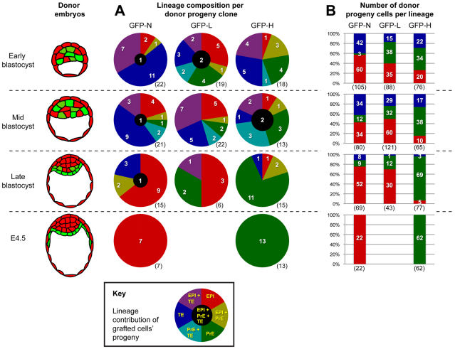
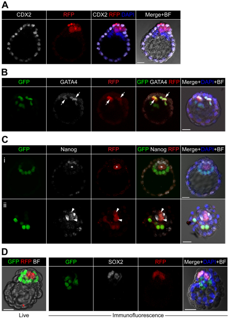
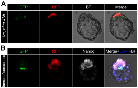
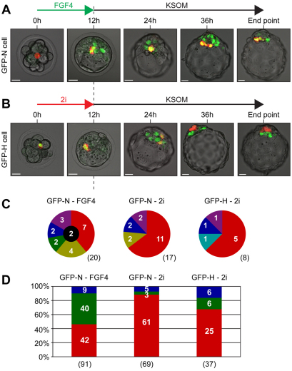
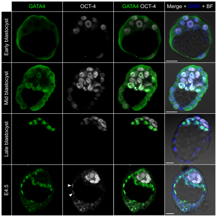
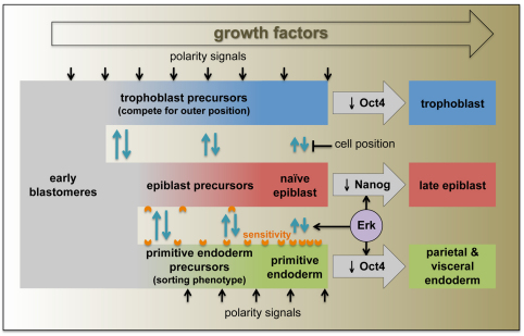
Cell differentiation during pre-implantation mammalian development involves the formation of two extra-embryonic lineages: trophoblast and primitive endoderm (PrE). A subset of cells within the inner cell mass (ICM) of the blastocyst does not respond to differentiation signals and forms the pluripotent epiblast, which gives rise to all of the tissues in the adult body. How this group of cells is set aside remains unknown. Recent studies documented distinct sequential phases of marker expression during the segregation of epiblast and PrE within the ICM. However, the connection between marker expression and lineage commitment remains unclear. Using a fluorescent reporter for PrE, we investigated the plasticity of epiblast and PrE precursors. Our observations reveal that loss of plasticity does not coincide directly with lineage restriction of epiblast and PrE markers, but rather with exclusion of the pluripotency marker Oct4 from the PrE. We note that individual ICM cells can contribute to all three lineages of the blastocyst until peri-implantation. However, epiblast precursors exhibit less plasticity than precursors of PrE, probably owing to differences in responsiveness to extracellular signalling. We therefore propose that the early embryo environment restricts the fate choice of epiblast but not PrE precursors, thus ensuring the formation and preservation of the pluripotent foetal lineage.


Figures

Fig. 1.
Fig. 1.
Progeny of ICM cells contribute to epiblast, PrE and trophoblast lineages when aggregated with morulae. (A) Experimental scheme. ICM cells from donor embryos were obtained by immunosurgery and disaggregation, selected on intensity of GFP signal and reaggregated with recipient morulae. (B) Examples of clonal contribution of donor cells in chimaeras after 48 hours in culture (end point): (i) epiblast clone; (ii) PrE clone; (iii) trophoblast clone; (iv) epiblast + PrE clone; (v) epiblast + trophoblast clone; (vi) tri-lineage clone. Arrows indicate a trophoblast cell; arrowheads indicate a PrE cell; asterisks indicate an epiblast cell. Scale bars: 20 μm.
Fig. 2.
Fig. 2.
Profile of lineage contribution of transplanted cells in experimental groups. (Left) schematic representation of blastocyst stages from which donor cells were isolated. (A) Proportions of donor cells contributing to specific combinations of lineages. (B) Proportions of donor cell progeny contributing to each lineage. Absolute numbers are indicated in white; sample sizes are in parentheses.
Fig. 3.
Fig. 3.
Expression of lineage markers in chimaeras. Donor progeny clones visualised as RFP-expressing (red) cells. Green fluorescence represents expression of PdgfraH2B-GFP. (A) Cdx2 expression in a clone contributing to both epiblast and trophoblast. All trophoblast cells are positive for Cdx2, although the level of expression differs. Epiblast cells are all negative for Cdx2. (B) Gata4 expression in a clone contributing only to PrE. Gata4 is present in all PrE cells and overlaps with expression of GFP (arrows). (C) Two examples of Nanog expression in chimaeras. (i) GFP-N donor progeny contributed only to epiblast, in which Nanog is absent in donor progeny (asterisk) but present in recipient epiblast (GFP negative and RFP negative) cells. (ii) GFP-H donor progeny contributed to both epiblast and PrE. Nanog is present in donor cells in the epiblast part of the clone (GFP negative and RFP positive, arrowheads), but not in donor cells in the PrE part of the clone (GFP positive and RFP positive). (D) Example of Sox2 expression in chimaera where GFP-N donor progeny contributed to epiblast. First on the left is a live image of the same embryo after 48 hours of culture. Scale bars: 20 μm.
Fig. 4.
Fig. 4.
Donor cells in chimaeras treated with inhibitors contribute solely to epiblast and express Nanog. (A) Live chimaeric embryo after 48 hours of culture. (B) Expression of Nanog visualised by immunofluorescence. Donor progeny are RFP expressing. GFP represents expression of PdgfraH2B-GFP. Scale bars: 20 μm.
Fig. 5.
Fig. 5.
Early transient FGF4 or 2i treatment influences lineage contribution of donor progeny. (A) Time-lapse imaging of chimaera from early GFP-N donor cell treated with FGF4 for 12 hours until early blastocyst formation. (B) Time-lapse imaging of chimaera from early GFP-H donor cell treated with 2i for 12 hours until early blastocyst formation. (C) Lineage contribution per donor progeny clone for each treatment. (D) Number of donor cell progeny per lineage for each treatment. Absolute numbers are indicated in white; sample sizes are in parentheses. Scale bars: 20 μm.
Fig. 6.
Fig. 6.
Expression of Oct4 and Gata4 in donor littermates. In early and mid blastocysts, Oct4 is expressed throughout the ICM and more weakly in trophoblast cells, whereas Gata4 is absent. From the mid blastocyst stage, Gata4 is detected in nuclei of the nascent PrE layer and more weakly in some deeper cells. By the late blastocyst, Gata4 is restricted to the PrE layer, in which Oct4 staining is weaker than in the epiblast and is fully restricted to the epiblast by E4.5 (arrowheads). Scale bars: 20 μm.
Fig. 7.
Fig. 7.
Model for changing cell states during pre-implantation development. Epiblast precursors, characterised by Nanog expression, represent a naïve totipotent state that can convert to primed PrE precursors in response to Erk activation. The properties of PrE precursors predisposes them to cell sorting, after which positional signals reinforce commitment to a PrE fate. Likewise, eventual loss of Nanog in epiblast precursors, which are protected from differentiation signals by their inside position, leads to epiblast commitment. We speculate that, in the absence of Erk activation, naïve epiblast precursors can also pass through a trophoblast precursor state that becomes reinforced by positional signals.

References

    1. Arman E., Haffner-Krausz R., Chen Y., Heath J. K., Lonai P. (1998). Targeted disruption of fibroblast growth factor (FGF) receptor 2 suggests a role for FGF signaling in pregastrulation mammalian development. Proc. Natl. Acad. Sci. USA 95, 5082–5087 - PMC - PubMed
    1. Brons I. G., Smithers L. E., Trotter M. W., Rugg-Gunn P., Sun B., Chuva de Sousa Lopes S. M., Howlett S. K., Clarkson A., Ahrlund-Richter L., Pedersen R. A., et al. (2007). Derivation of pluripotent epiblast stem cells from mammalian embryos. Nature 448, 191–185 - PubMed
    1. Chambers I., Colby D., Robertson M., Nichols J., Lee S., Tweedie S., Smith A. (2003). Functional expression cloning of Nanog, a pluripotency sustaining factor in embryonic stem cells. Cell 113, 643–655 - PubMed
    1. Chawengsaksophak K., de Graaff W., Rossant J., Deschamps J., Beck F. (2004). Cdx2 is essential for axial elongation in mouse development. Proc. Natl. Acad. Sci. USA 101, 7641–7645 - PMC - PubMed
    1. Chazaud C., Yamanaka Y., Pawson T., Rossant J. (2006). Early lineage segregation between epiblast and primitive endoderm in mouse blastocysts through the Grb2-MAPK pathway. Dev. Cell 10, 615–624 - PubMed

Publication types

MeSH terms

Substances