. 2012 Jan;139(1):33-45.
doi: 10.1242/dev.071761. Epub 2011 Nov 17.

Ptf1a-mediated control of Dll1 reveals an alternative to the lateral inhibition mechanism

Affiliations

Ptf1a-mediated control of Dll1 reveals an alternative to the lateral inhibition mechanism

Jonas Ahnfelt-Rønne et al. Development. 2012 Jan.

Abstract

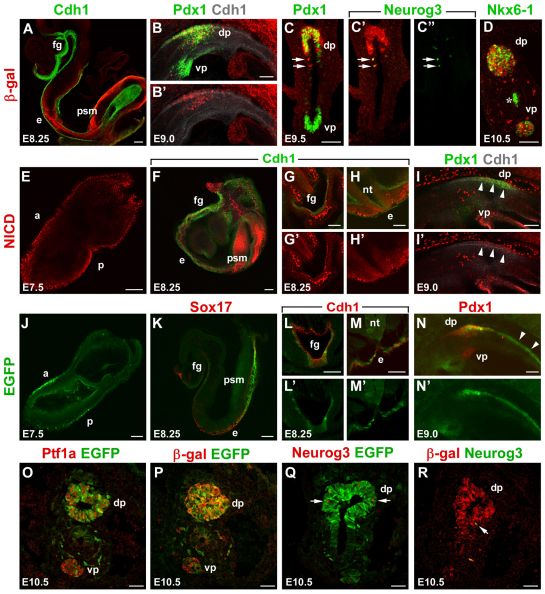
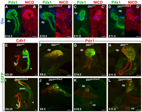
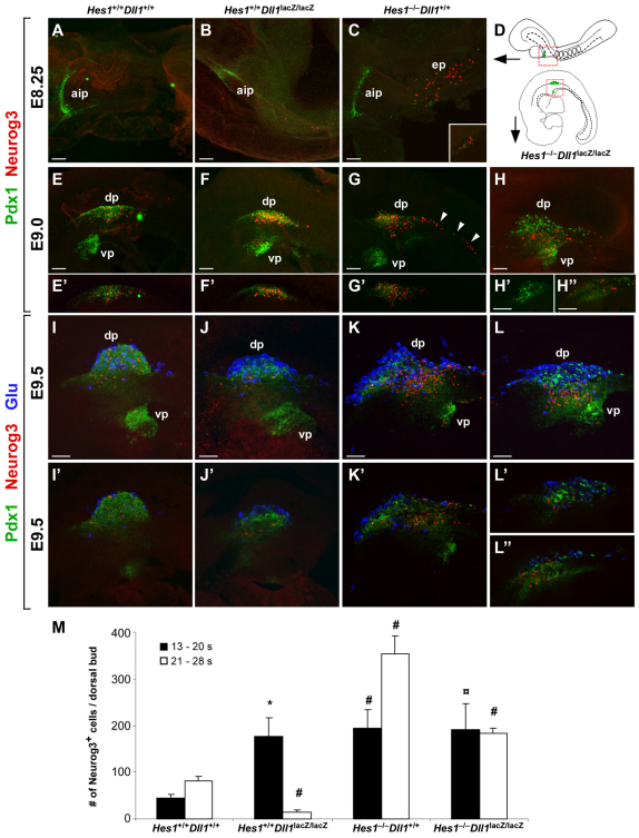
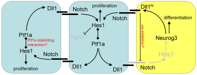
Neurog3-induced Dll1 expression in pancreatic endocrine progenitors ostensibly activates Hes1 expression via Notch and thereby represses Neurog3 and endocrine differentiation in neighboring cells by lateral inhibition. Here we show in mouse that Dll1 and Hes1 expression deviate during regionalization of early endoderm, and later during early pancreas morphogenesis. At that time, Ptf1a activates Dll1 in multipotent pancreatic progenitor cells (MPCs), and Hes1 expression becomes Dll1 dependent over a brief time window. Moreover, Dll1, Hes1 and Dll1/Hes1 mutant phenotypes diverge during organ regionalization, become congruent at early bud stages, and then diverge again at late bud stages. Persistent pancreatic hypoplasia in Dll1 mutants after eliminating Neurog3 expression and endocrine development, together with reduced proliferation of MPCs in both Dll1 and Hes1 mutants, reveals that the hypoplasia is caused by a growth defect rather than by progenitor depletion. Unexpectedly, we find that Hes1 is required to sustain Ptf1a expression, and in turn Dll1 expression in early MPCs. Our results show that Ptf1a-induced Dll1 expression stimulates MPC proliferation and pancreatic growth by maintaining Hes1 expression and Ptf1a protein levels.


Figures

Fig. 1.
Fig. 1.
Dll1, NICD and Hes1 expression in endoderm. (A-D) Optical sections of E7.5 to E10.5 Dll1lacZ/+ embryos whole-mount stained for β-galactosidase, Cdh1, Pdx1, Neurog3 or Nkx6-1. The arrows in C point to Neurog3+ cells that co-express β-gal. The asterisk in D marks trapping of the anti-Nkx6-1 antiserum in the gut lumen. (E-I′) Optical sections of E7.5 to E9.0 wild-type embryos whole-mount stained for NICD and Cdh1. The arrowheads in I and I′ indicate the NICD expression in the dorsal pancreatic epithelium. (J-N′) Optical sections of E7.5 to E9.0 Tg(Hes1-EGFP)1Hri embryos whole-mount stained for EGFP, Sox17, Cdh1 or Pdx1. The arrowheads in N point out Hes1-EGFP expression in the dorsal gut tube endoderm. (O-R) Cryosections of E10.5 Dll1lacZ/+ Tg(Hes1-EGFP)1Hri double heterozygotes (O,P), Tg(Hes1-EGFP)1Hri (Q) and Dll1lacZ/+ (R) embryos. (O) Double IF detection of Ptf1a and EGFP. (P) Double IF detection of β-galactosidase and EGFP. (Q) double IF detection of Neurog3 and EGFP. (R) Double IF detection of β-galactosidase and Neurog3. The arrows in Q and R point to Neurog3+ cells. a, anterior; dp, dorsal pancreas; e, endoderm; fg, foregut; nt, neural tube; p, posterior; psm, presomitic mesoderm; vp, ventral pancreas. Scale bars: 50 μm. See also supplementary material Fig. S1.
Fig. 2.
Fig. 2.
NICD and Hes1 expression in Dll1 mutants. (A-D) Optical sections of whole-mount IF-stained E10.5 and E11.5 Dll1+/+ and Dll1lacZ/lacZ embryos. The embryos were stained for Pdx1, NICD and glucagon. Note that NICD is reduced in E10.5 Dll1lacZ/lacZ embryos but recovers at E11.5. (E-L) Optical sections of Tg(Hes1-EGFP)1Hri mice on Dll1+/+ (E-H) or Dll1lacZ/lacZ (I-L) background. Endodermal EGFP expression is normal in E8.25 Dll1lacZ/lacZ mice but becomes reduced beginning at E9.5 and is essentially lost at E10.5. However, EGFP expression recovers at E11.5 coincident with NICD recovery. dp, dorsal pancreas; e, endoderm; fg, foregut; psm, presomitic mesoderm; vp, ventral pancreas. Scale bars: 50 μm.
Fig. 3.
Fig. 3.
Ptf1a is required for Dll1 expression in MPCs. (A-F′) Image stack projections (A-C) and optical sections (D-E) of whole-mount stained E10.5 embryos of the indicated genotypes, stained for β-galactosidase indicating Dll1 expression, Pdx1 and glucagon. A-C show triple labeled projections. A′-C′ shows red channel alone for an unobstructed view of β-gal expression. D-F and D′-F′ show optical sections of red/green and blue/green channels, respectively, of the embryos shown in A-C. dp, dorsal pancreas; vp, ventral pancreas. Scale bars: 50 μm. (G) ChIP-Seq data obtained with anti-Ptf1a antibodies (red line) and anti-Rbpj antibodies (blue line) with E15.5 chromatin and aligned with a multiple sequence alignment of the Dll1 5′-flanking region. Note that Ptf1a and Rbpj bind to conserved sequences in the 5′-flank. Nucleotide positions on chromosome 17 are indicated. Note that the Dll1 locus depicted in G is on the lower strand and hence reads right to left. See also supplementary material Fig. S2.
Fig. 4.
Fig. 4.
Dll1 is required for continued formation of endocrine precursors. (A-C) Image stack projections of E8.25 wild-type, Dll1 and Hes1 mutant embryos whole-mount stained for Pdx1 and Neurog3. The inset in C shows an optical section of emerging Pdx1+ cells in the dorsal endoderm. Note that A and C are ventral views, whereas B is tilted more laterally. (D) Schematics of E8.25 and E9.0-9.5 embryos with red boxes indicating imaged areas in the other panels. (E-H) Image stack projections of E9.0 wild-type, Dll1, Hes1 and Hes1/Dll1 mutant embryos whole-mount stained for Pdx1 and Neurog3. The arrowheads in G point to ectopic expression of Neurog3 in the dorsal gut epithelium posterior to the Pdx1+ pancreas area. (E′-H′) Optical sections through the dorsal bud region of the embryos shown in E-H. (H′) optical section through the lateral gut wall. (I-L) Image stack projections of E9.5 wild-type, Dll1, Hes1 and Hes1/Dll1 mutant embryos whole-mount stained for Pdx1, Neurog3 and glucagon. (I′-L′) Optical sections through the dorsal bud region of the embryos shown in I-L. (L′) Optical section through the lateral gut wall. aip, anterior intestinal portal; ep, endocrine progenitors; dp, dorsal pancreas; vp, ventral pancreas. Scale bars: 50 μm. (M) Bar graph showing quantification of Neurog3+ cells in E9.0 (13-20 somite) and E9.5 (21-28 somite) wild-type, Dll1, Hes1 and Hes1/Dll1 mutant embryos. Data are represented as mean ± s.d. #, P<0.001; *, P<0.002; formula image
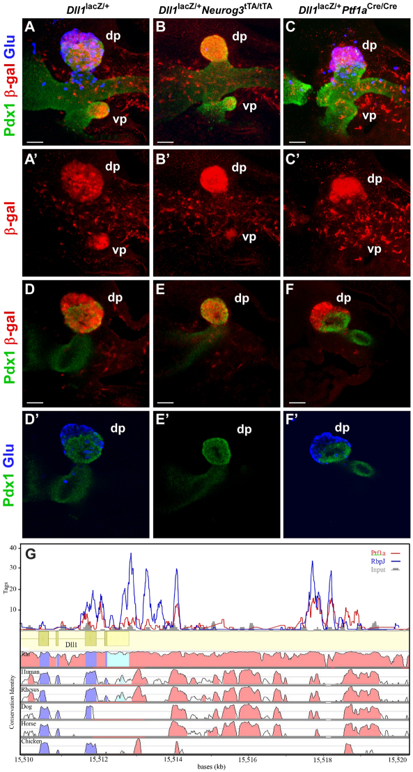
, P<0.005; by Student’s t-test (compared with wild type); n=3-9. See also supplementary material Fig. S3.
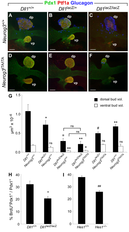
Fig. 5.
Fig. 5.
Ptf1a-mediated Dll1 expression is required for MPC proliferation. (A-F) Image stack projections of E10.5 embryos from crosses of Dll1lacZ/+Neurog3tTA/+ double-heterozygote animals, whole-mount stained for Pdx1, Ptf1a and glucagon. Note that dorsal and ventral bud size is equally reduced in Dll1lacZ/lacZNeurog3+/+ and Dll1lacZ/lacZNeurog3tTA/tTA embryos. dp, dorsal pancreas; vp, ventral pancreas. Scale bars: 50 μm. (G) Quantification of dorsal and ventral bud volume in wild-type and mutant embryos of the indicated genotypes based on Pdx1/Ptf1a double whole-mount stained and confocally scanned embryos. (H,I) Quantification of BrdU incorporation in E10.5 Pdx1+ MPCs in Dll1 and Hes1 mutant dorsal buds, respectively. Data in G-I are represented as mean ± s.d. ##, P<0.0002; formula imageformula image, P<0.0005; #, P<0.002; formula image, P<0.005; **, P<0.01; *, P<0.05; by Student’s t-test (compared with wild type unless otherwise indicated); n=2-4. ns, not significant.
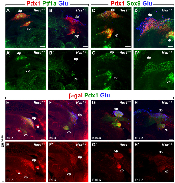
Fig. 6.
Fig. 6.
Hes1 is required for normal Ptf1a and Dll1 expression in MPCs. (A-D′) Image stack projections of whole-mount stained E9.5 wild-type (A,C) and Hes1–/– (B,D) embryos, showing expression of Pdx1, glucagon and Ptf1a (A,B) or Sox9 (C,D). A′-D′ show the green channel alone to give an unobstructed view. Note the loss of Ptf1a immunoreactivity in Hes1–/– dorsal bud. (E-H′) Image stack projections of whole-mount stained E9.5 (E,F) and E10.5 (G,H) wild-type (E,G) and Hes1–/– (F,H) embryos, showing expression of Dll1 (β-gal), Pdx1 and glucagon. E′-H′ show the red channel alone to give an unobstructed view. Note the reduced levels of β-gal expression in E9.5 and E10.5 dorsal buds and E10.5 ventral buds in Hes1 mutants. The asterisks in E mark specks of non-specific fluorescence from the secondary antibody. dp, dorsal pancreas; vp, ventral pancreas. Scale bars: 50 μm. See also supplementary material Fig. S4.
Fig. 7.
Fig. 7.
Model of the relationships between transcription factors and Notch signaling components in primary transition pancreas. In contrast to the simple lateral inhibition model, the new model features Ptf1a-mediated activation of Dll1, which is crucial for Notch activation in MPCs. Furthermore, the model proposes that Notch is subject to cis-inhibition by high levels of Dll1 induced by Neurog3. Activated components of the regulatory system are shown in black, whereas components held inactive are gray.

References

    1. Ahnfelt-Ronne J., Hald J., Bodker A., Yassin H., Serup P., Hecksher-Sorensen J. (2007a). Preservation of proliferating pancreatic progenitor cells by Delta-Notch signaling in the embryonic chicken pancreas. BMC Dev. Biol. 7, 63 - PMC - PubMed
    1. Ahnfelt-Ronne J., Jorgensen M. C., Hald J., Madsen O. D., Serup P., Hecksher-Sorensen J. (2007b). An improved method for three-dimensional reconstruction of protein expression patterns in intact mouse and chicken embryos and organs. J. Histochem. Cytochem. 55, 925–930 - PubMed
    1. Apelqvist A., Li H., Sommer L., Beatus P., Anderson D. J., Honjo T., Hrabe de Angelis M., Lendahl U., Edlund H. (1999). Notch signalling controls pancreatic cell differentiation. Nature 400, 877–881 - PubMed
    1. Beres T. M., Masui T., Swift G. H., Shi L., Henke R. M., MacDonald R. J. (2006). PTF1 is an organ-specific and Notch-independent basic helix-loop-helix complex containing the mammalian Suppressor of Hairless (RBP-J) or its paralogue, RBP-L. Mol. Cell. Biol. 26, 117–130 - PMC - PubMed
    1. Bettenhausen B., Hrabe de Angelis M., Simon D., Guenet J. L., Gossler A. (1995). Transient and restricted expression during mouse embryogenesis of Dll1, a murine gene closely related to Drosophila Delta. Development 121, 2407–2418 - PubMed

Publication types

MeSH terms